Going beyond faked data… faking the raw data!

Most readers here will be familiar with the issues surrounding western blotting as a technique in the bio-sciences, namely it is highly amenable to fabrication and inappropriate manipulation. A quick glance at PubPeer, ScienceIntegrityDigest, ForBetterScience, and other blogs reporting on scientific misconduct, makes it clear that blot-fakery is alive and well in 2020. Today, I will walk you through a paper I received for review which takes this to a whole new level!

What’s the problem?

One of the most common problems encountered with western blot data, is the presentation of “letter-boxed” blot images such as the one shown here…

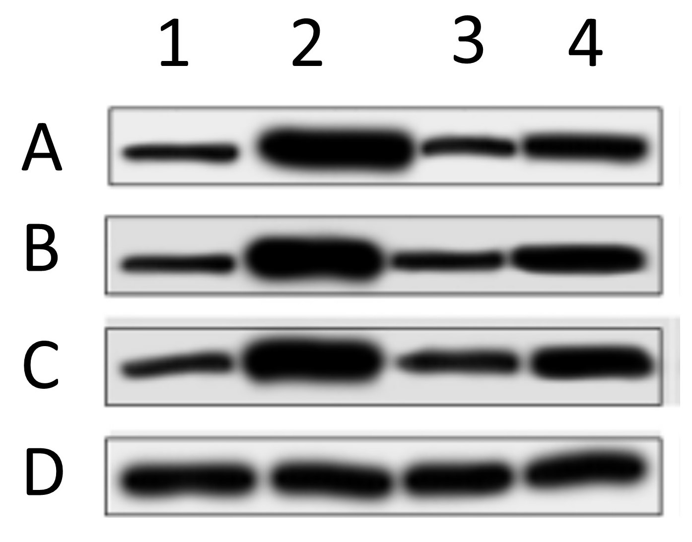
In this image, 4 different proteins (ABCD) are blotted under 4 different experimental conditions (1234). These types of image are problematic for several reasons. First, there are no molecular weight (MW) markers, so it’s impossible to tell if the antibody recognized a protein at the correct MW. Second, only a small vertical slice of gel is shown, so it’s impossible to tell if the antibody only recognized one protein or several bands (how specific was the Ab). Third, in these types of collages the bottom blot (D in this case) is typically the “loading control”, showing the abundance of a house-keeping protein such as GAPDH or beta-actin, thus allegedly proving that equal protein was loaded in each lane. The problem is, as you can see from the example above, the spacing, size, shape, and slope of the bands is different in each blot. This makes it clear that the “loading control” likely did not originate from the same blot membrane – the authors simply loaded a different gel and blotted that, so this is not a true loading control. Fourth, western blotting as a method suffers from a woefully narrow dynamic range (about 10-fold), so it’s important for quantitation purposes that the bands be exposed somewhere within that range. Here, all of the bands have solid black centers and are overexposed, so the bands are completely useless for quantitative purposes. Lastly, the background (more on that in a minute) is plain and washed out, so it’s impossible to “anchor” the bands to any background features. This makes it impossible to tell if any of the bands have been pasted or spliced together. It doesn’t mean the image is fake, it just means it’s impossible to say it’s not fake.

When I receive a paper to review, if the bulk of data is presented in this letter-boxed format, I will often reject the manuscript, or demand better quality evidence.

How have publishers reacted to the blot-fakery crisis?

In recent years, many journals have become aware of the shit show that is blot-fakery, and have started to demand better quality controls for submitted manuscripts. This typically requires provision of full-sized, uncropped blots as original images. Since most journals are online and storage is not an obstacle, this is now easy for authors (of authentic work) to comply with. Many publishers are going a step further and demanding complete original data sets behind every figure! My lab has been doing this for a while now, posting complete data sets on the file server FigShare.

How have faking authors responded?

One might think that simply demanding better quality original data would solve the blot-faking crisis, but no. What if the “original” blot images can be faked? That’s what I found today while reviewing a paper for a UK based journal.

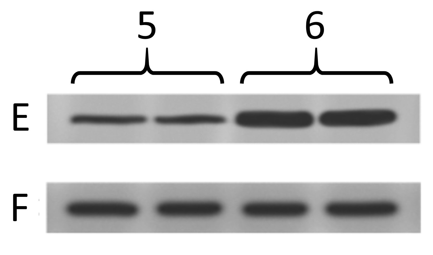
Below are the two blots as presented in the paper. The one on the left is the same as at the top of this post, and was from cells treated with 4 different drug conditions. The other one is from cells treated with 2 different genetic manipulations (5, 6) and blotting 2 more proteins (E, F) where F is the “loading control”.

Now, below are the “original” blot images whence (allegedly) these letterboxed blots came. On the left, the 4 blots that make up the first panel, and on the right the 2 that make up the second panel. At first glance, this seems like the epitome of data transparency; beautiful full-sized gel images with lovely backgrounds, and band patterns that clearly match up to the images used in the panels for the final paper.

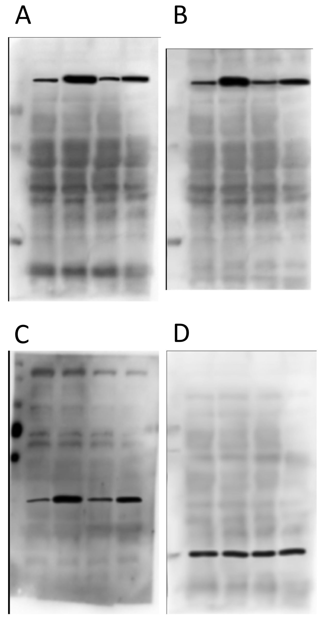
So what’s wrong? Let’s take a look at panels A and B first…

As you can see from the red boxes and the blue arrows, there are several features shared between these 2 background images. What makes this interesting is that protein A and protein B are of a very similar molecular weight – it’s quite common to strip and re-probe a blot using a different antibody, or to cut up a membrane and probe for several proteins at once, but to blot for 2 proteins at an almost identical molecular weight is almost impossible to pull off. The other weird thing is that the horizontal width of some of the bands appears to have been adjusted between these blot background images (e.g. the material in the 3rd lane appears wider in B than in A, as if it’s been stretched).

Next up, proteins C and D from the first panel…

There’s nothing particularly egregious about this pair. It is clear they’re from different gels/membranes due to the different backgrounds, which is a good thing. BUT… the protein in D is the “loading control”, and so is meant to come from the same membrane. Ergo, this is not a true loading control – they just loaded the same samples on a different gel. Lots of people do this and it’s not good.

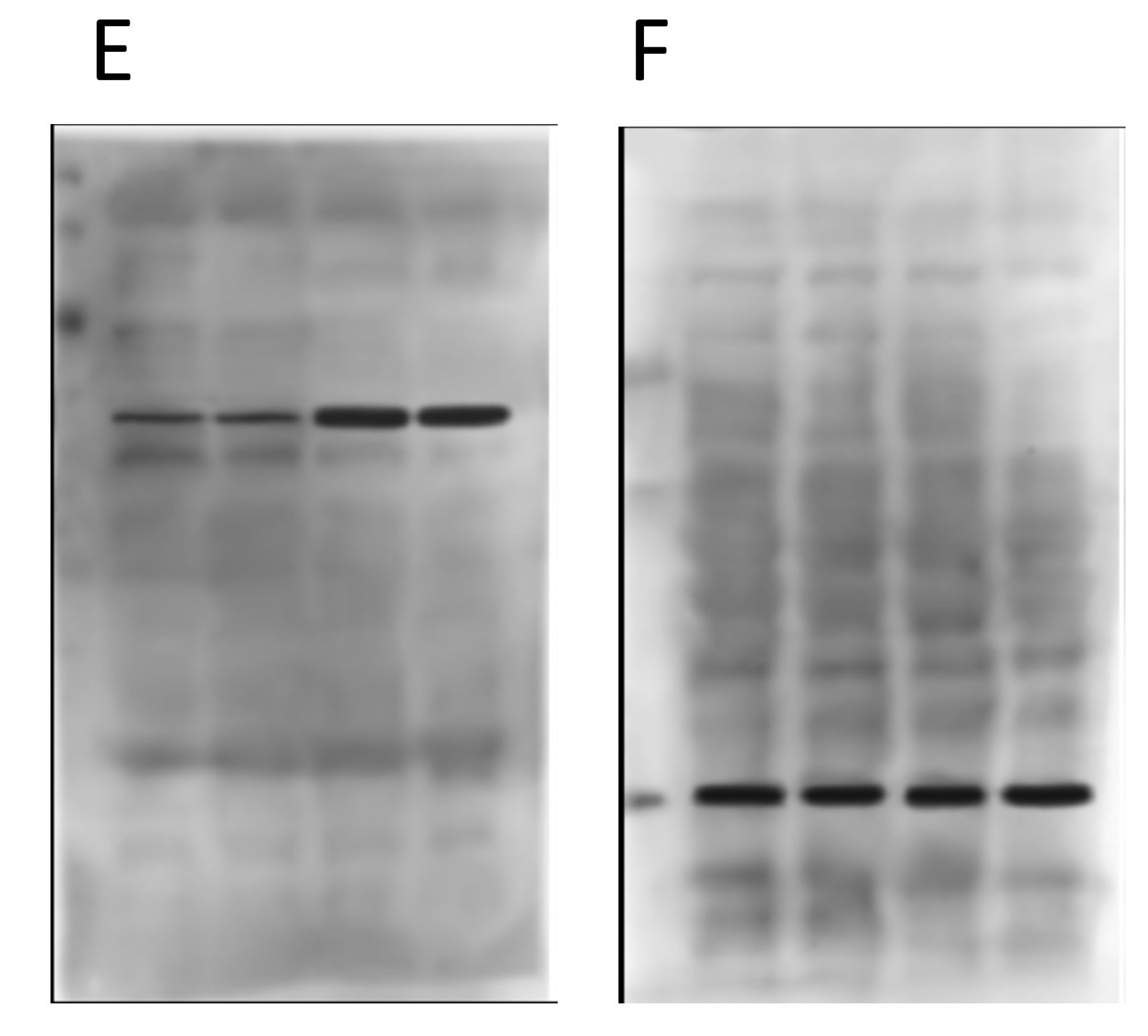
Now onto the proteins E and F in the second panel…

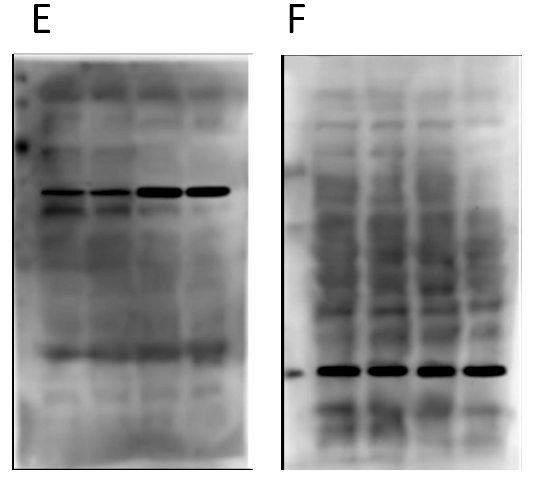
Again, nothing particularly egregious here. Different proteins, different blot images, different backgrounds. But again, F is the loading control protein, so they should have been blotted/probed on the same membrane. Not using proper “loading controls” doesn’t adhere to best practices.

So what’s the big deal?

Things start to get real interesting when we compare the backgrounds of the “original” blot images from the two different panels. Remember, in the original paper the 4 blots A/B/C/D were from cells treated with drugs, and the 2 blots E/F were from genetically manipulated cells. Completely different experiments – no way the blots can be the same right? Right…

Obviously there’s been some re-sizing, and they’re not identical, but as the colored boxes show there are a number of very coincidental similarities between these two background images, onto which the black bands for the proteins of interest seem to have been teleported. There are also some funny shadow lines above the black band of interest in the right hand image (E) which may be indicative of splicing.

Furthermore, when we compare the images for panels D and F, i.e. the two loading controls from the completely different and unrelated experiments, there are also common features on the blot backgrounds…

There are differences, but there are more similarities than would be expected by chance, if these two “original blot images” really originated from completely different experiments.

So what can we do about it?

First to summarize, a detailed analysis of the backgrounds for the “original blot images” provided for this paper does not instill any confidence in the integrity of the data. It appears as if the proteins of interest (solid black bands) have been pasted onto background images, to “generate” original blot images.

In terms of what can be done about this type of data fakery, one answer is posts like this, to highlight the problem to journal editors. Even in our new-found utopia of data transparency and open availability of “original” data, authors continue to dupe reviewers and editors, so we need to be increasingly vigilant.

Another solution is to name and shame. Unfortunately this would be problematic on several counts. The journal review process is private, and if I were to reveal the name of the journal they would probably demand I take down the above images since they were provided to me in confidentiality. For this reason, I removed any identifying labels about the proteins. It would also not be particularly fair on the authors for their work to be thrown into the court of public opinion before it has had a chance for proper peer-review.

In this case, I rejected the paper and I outlined the reasons why in my review. But, as is usually the case, I expect it will eventually show up published in another journal. This has happened a few times… I call out a paper during review and it shows up later with the offending data removed (or sometimes not) but with the same list of authors, thus indicating the senior author did not wish to punish whomever in their lab did the faking.

So, for now I’ll be watching closely to see if this paper makes an appearance somewhere else, and (naturally) I’ll be paying close scrutiny to the other papers from this group, to see if there are any other shenanigans worth reporting here or on PubPeer. All of this just goes to show that even with enhanced data stewardship approaches, the plague of western blot fakery shows no signs of going away.

ALKBH7

This post is a bit late, but I wanted to record the story behind our recent eLife paper on ALKBH7 somewhere less ethereal than Twitter.

The story for us started as a collaboration with Dragony Fu in UofR’s Department of Biology. Dragony has worked on ALKBH7 for a number of years, and had shown that it plays a critical role in programmed necrosis in response to DNA alkylation. In addition there was some earlier work showing that the Alkbh7-/- mouse was obese and had some fat oxidation problems.

ALKBH7 is a mitochondrially-localized member of the alpha-ketoglutarate (aKG) dependent dioxygenase family. This includes enzymes you’ve probably heard of such as the TETs, the Jumonji domain demethylases, and the EGLN family of prolyl hydroxylases that regulate HIF. All these proteins add -OH onto something, for varied reasons. For example the DNA demethylases perform a “demethylation via hydroxylation” fundtion – they add -OH to the methyl group, which then spontanously decomposes, giving back the original non-methylated DNA base and formaldehyde. ALKBH7 is a homolog of the E. coli DNA repair enzyme ALKB, and pretty much all the other members are involved in repairing DNA alkylation damage.

The problem is, nobody has ever found a substrate for ALKBH7! It lacks the usual DNA binding pocket that other ALKB proteins contain. In-fact, the only thing it’s ever been shown to do is hydroxylate itself on a leucine. So, we hypothesized that ALKBH7 might be a mitochondrial prolyl-hydroxylase. We sent a bunch of WT and KO heart samples off to ‘Tish Murphy’s lab, where Leslie Kennedy performed a proteomic analysis – the idea was that if ALKBH7 is a prolyl-hydroxylase, we should see less P-OH in its substrate proteins, in the knockout.

Nothing! No differences. Well, there was one protein that showed lower hydroxylation… hydroxyacyl-CoA dehydrogenase. This was interesting at first, because remember the knockouts are obese. What if prolyl-hydroxylation was a novel mechanism of regulating fatty acid beta-oxidation? Well we did a bunch of enzymology on the dehydrogenase and nothing panned out, so that was a dead end.

As part of the proteomic analysis, we also had an abundance data set, giving us the levels of 3700 proteins in the WT vs. KO hearts, and here’s where things got interesting… only 2 proteins were up, and one of them was glyoxylase I (GLO-1). We confirmed this both by western blot and by doing GLO-1 activity assays, and the effect was real.

So, what’s GLO-1? (it’s also worth noting we ignored this finding for several months because the protein was listed as “lactoylglutathione lyase” in the data set, and we didn’t know what the hell that was all about, so…) Anyway, GLO-1 is a key enzyme involved in the detoxification of methylglyoxal (MGO), which is a toxic byproduct of glycolysis. Excess glucose metabolism such as occurs in hyperglycemia and diabetes, leads to more MGO, which can react with various biomolecules to form “advanced glycation end products” (AGEs). These post-translational modifications essentially gum up protein fucntion, and this is what’s believed to drive a lot of the pathology of diabetes. When a diabetic patient gets tested for glycated hemoglobin (aka “HbA1C” or simply A1C), that’s the MGO adducted form of hemoglobin, used to indicate long term trends in blood glucose. We also did a bunch of metabolomics analysis, to show that glycolysis is up in the ALKBH7 knockouts.

So, what does any of this have to do with cardiac ischemia-reperfusion (IR) injury, the main thing that we study? Well, the main mode of cell death in IR injury is necrosis, and remember ALKBH7 is required for necrosis. So, sure enough, we were able to show that the Alkbh7-/- mouse is protected against cardiac IR injury. We also made an inhibitor for ALKBH7 and showed that it is protective too. AND we showed that blocking the GLO-1 enzyme prevents the protection in the Alkbh7-/- mice. So, GLO-1 is required for the cardioprotection.

For most who study mitochondria and cardiac IR injury or protection, the mitochondrial permeability transition (PT) pore is the go-to target when we find an effect. But, we measured PT pore opening in Alkbh7-/- and it just wasn’t very impressive. Sure, there was a small change, but nowhere near enough to protect the heart from IR injury. So we struck out again. We also had recently shown that the induction of the mitochondrial unfolded protein response (UPRmt) was capable of inducing protection, but again no differences seen in the UPRmt in Alkbh7-/- mice. More negative data!

So, the upshot of all this is that we STILL don’t really understand how ALKBH7 is required for necrosis in heart attack. When you knock out ALKBH7, there’s an upregulation of GLO-1 and a rewiring of all the bits of metabolism that make MGO, and that appears to be required for the protection. But exactly how this protein in the mitochondrion signals to MGO production in the cytosol (where glycolysis is), is still not well understood.

Of course, the final writing and revision of the paper took place during the whole #Covid19 lock down and gradual reopening process, which essentially killed our ability to take a deeper dive and really close out the story. At the end of the day, it’s hard to study an enzyme for which there is no known activity and no known substrate! We’re still working in this area, and hope to be able to address some of these unknowns soon (for example by developing a screening assay for potential ALKBH7 substrates).

In sum, this started out as a collaboration with a biology colleague, took in a multi-omics approach (proteomics, metabolomics, PTM proteomics) and a bunch of other methods (the paper has >60 panels of data), and frankly most of what we found was negative. It’s frustrating, but that’s sometimes how science works. The good thing is we learned a bunch of interesting stuff along the way, and that brings us closer to understanding cardiac metabolism and how it can be manipulated for therapeutic benefit in situations such as IR.

As is usual for us now, the paper was posted as a preprint on @BioRxiv, and we also posted full data sets (humongous proteomics files) on FigShare.

#COVID19 Lab non-happenings

So, like most of the world, the lab has been in lock-down since early March (we ramped everything down, including culling our mouse colonies to 1/3 of their original size, and turned off the lights on March 12th). Nevertheless, things have been moving along…

(1) Work with our collaborator Sabzali Javadov at University of Puerto Rico was published. along with an editorial for AJP Lung written with Mike O”Reilly, all about Scott Ballinger’s trans-mitochondrial mouse study.

(2) Lots of grants submitted and reviewed… In February we submitted an NIDDK R01 on the potential role of mitochondrial K+ channels as anti-obesity drug targets, as well as a metabolomics collaboration with Heiko Bugger in Austria. Paul served on an NIH special emphasis panel, and will also be ad-hocking (sp?) for NIH in June.

(3) Our mass spec’ went kaput! Apparently either the turbo pump or its controller are dead, so we’re awaiting a service call to fix it, but of course with the virus shut-down it’s not clear when that will be accomplished.

(4) Thankfully, when the lock-down occurred, we were at a place in the research cycle where we’re sitting on a LOT of data, and so now we’re writing up several papers for submission in a few weeks. While (my guess is) the rest of the world is going to be churning out review articles during this time, I’m hopeful that we can get some actual science published this year!

(5) We were intrigued by the report last fall from Yingming Zhao’s group regarding the potential for modification of histone lysine residues by “lactylation” (addition of lactate)… especially since, at the same time, Jim Galligan’s group in Arizona reported a possible mechanism. However, there were some problems in the Zhao paper related to the anti-lactyl-lysine antibody, and we currently have in revision at Nature a “Brief Matters Arising” paper, outlining some important caveats. We may post a pre-print on BioRxiv in the coming weeks, depending on how fast things move through the editorial process.

As seems to be the case with so much scientific communication recently, the blog is going the way of the dodo, and Twitter seems to be where it’s at, for more timely updates on happenings of both a scientific and non-scientific nature.

Update on papers & other happenings

Some papers that have been in the pipeline for quite some time are now finally in the wild

(1) Our paper showing how cardio-protection by the mitochondrial unfolded protein response (UPRmt) depends on the transcription factor ATF5, is now out in AJP Heart.

(2) From our long-running collaboration with Keith Nehrke, some work from his post-doc’ Yunki Im showing that the post-fertilization elimination of paternal mitochondria employs the FNDC1 mitophagy pathway. Published in Dev. Biol.

(3) From a collaboration with Paige Lawrence‘s lab, a paper in Scientific Reports on how early-life exposure to aryl-hydrocarbon receptor agonists has long-term impacts on mitochondrial function in T-Cells.

Other news –

Paige was also instrumental in the recent acquisition of a new Seahorse XFe96 analyzer for the URMC Shared Resource Laboratories. This came not a moment too soon, as our own ancient (serial # 003) XF24 machine died, and our XF96 will no longer be supported after next year.

PSBLAB grad’ student Alexander Milliken is about to do his qualifying exam in a couple of weeks, closely followed by former rotation student Jessica Ciesla (in the lab of Josh Munger).

As reported on Twitter, Alan Cash (the CEO of various corporations selling oxaloacetate as a supplement) called me up and threatened to sue over things written in this article. No letter yet.

We had a great time at the AHA BCVS meeting in Boston, catching up with old science friends and making new ones. Next travel is NHLBI mitochondria meeting in DC at the end of September, and then NIH MIM study section in DC in October.

Congratulations to our colleague George Porter, who got his R01 funded (on cyclophilin D and the PT pore in early cardiac development), and is now searching for a mitochondriac post-doctoral fellow.

Oxaloacetate Shenanigans

I was at a meeting yesterday discussing cancer treatments, and the use of oxaloacetate (OAA) as an anti-cancer drug came up. This prompted some more reading, and my conclusion is there’s a lot of craaaazy stuff out there, with very little actual evidence for pushing this molecule into the clinic.

Background on OAA

First, for the uninitiated, OAA is one of the metabolites in the Krebs’ tricarboxylic acid (TCA) cycle in mitochondria. OAA is made by malate dehydrogenase (MDH)…

The MDH reaction is energetically unfavorable (ΔG +29.7 kJ/mol), but in reality that’s overcome by the next reaction in the Krebs’ cycle (citrate synthase, CS) being very favorable (ΔG -31.5 kJ/mol) so OAA gets removed as soon as it is made and that pulls the cycle forward.

OAA as a Drug?

The development of OAA from metabolite to drug appears to be pushed mostly by a company called “Terra Biological“. They’re selling OAA as a nutraceutical dietary supplement (only $49 for a month’s supply!) with all of the usual snake-oil claims that accompany such operations (“boosts energy”, “enhances mental focus”, blah-di-blah). Oh, and it allegedly mimics caloric restriction (CR) so will help you live longer, so there’s that. Here‘s a slide deck by the CEO of the company, Alan B. Cash, pushing all kinds of uses of OAA, from cancer to Parkinson’s and Alzhemier’s disease and everything in between (and here‘s another link in case that one goes down). The company received a warning letter from the FDA in 2017, for making drug-like claims about OAA supplements.

Despite this craziness, the company is now pushing OAA into the disease area of Glioma, by promoting “Cronoxal” (aka OAA) as a “medical food” for glioma patients. This DropBox link contains a bunch of hokey information from the company on the alleged benefits. For a time, Cronoxal was also known as “Gliaxal”. It’s also marketed under the name Benagene a “genomic aging supplement” (whatever that means) and as Jubilance for PMS relief. Cash also runs MetVital, yet another company developing OAA in a bunch of disease areas.

So where’s the evidence?

As is often the case with the murky waters of poorly-regulated dietary supplements are a number of shortcomings with the actual underlying scientific evidence for OAA’s effects. Most of the hype appears to hinge on this 2009 paper in Aging Cell, claiming that addition of 8 mM (!) OAA to growth media enhances lifespan in the nematode C. elegans. A notable feature of this paper is that it was published in September 2009 and contains no conflict of interest statement.

Compare and contrast with an October 2009 review article (Submitted April that year) in the OA journal Open Longevity Science (which no longer exists), in which author Alan B. Cash lists a conflict of interest – namely being an officer of a company that sells OAA supplements (that would be Terra Biological, founded in 2006).

So, Dr. Cash (and the lead author on the Aging Cell paper David Williams) appear to have seen fit to disclose this rather massive COI in an OA review article, but not in an Aging Cell primary research paper. Another notable find is that Cash submitted a patent application on OAA as a CR mimetic in 2005 – a full 4 years before the paper was published. You might think such an event would require disclosure?

Back to the science, Cash’s glorious PowerPoint deck from 2014 includes claims that studies in mice were underway in 2011, in collaboration with Steve Spindler at UC Riverside. A quick PubMed search indicates that nothing was ever published on this. Similarly, claims are made about lifespan extension in flies, but again there’s nothing published.

For brain cancer, there are a couple of papers showing alleged effects of OAA in mice, but the data are just not very believable, and the dose used – 2 grams per kg (that’s not a typo) equating to 150 grams in a human – is ridiculous!

So, that leaves just about the entirety of the medical claims about OAA based on a single published paper about worms, a shady pay-to-play OA review article by the CEO of the company, and a bunch of unsubstantiated and unpublished claims in PowerPoint slides. Despite this, some people saw fit to convince the FDA to allow clinical trials – one for Parkinson’s, which failed, and another for Alzheimer’s which is ongoing (wanna bet how it’ll turn out?)

OAA cannot work the way they say it does

The claimed mechanism-of-action for OAA is that it drives the MDH enzyme reaction backwards, and this enhances levels of NAD+, which boosts the activity of the Sirtuins, which are allegedly involved in the beneficial effects of CR. There’s another whole can of worms about SIRTs and NAD+ (covered briefly here), but that’s for another day. But anyway, driving MDH backwards to make NAD+ has a number of problems…

First, by using this redox reaction as a source of NAD+, the ultimate source is of course NADH. In cells, NAD+ is normally present at about 700-fold greater levels than NADH. As such, NADH cannot possibly serve as an adequate “reservoir” to provide more NAD+. Even if you could convert all the NADH in your cells to NAD+, the effective level of NAD+ would only go up by 0.14% (and you’d probably die because – duh – no NADH left!)

OAA (wikipedia)

Second, OAA (above) is a dicarboxylic acid. As such, it is negatively charged at physiologic pH. Negatively charged species are generally excluded from cells. Any mitochondrial biologist can tell you the way to get dicarboxylates into cells is to block the carboxylate moieties with alkyl groups… You make methyl or ethyl esters and these get transported into cells, then the alkyl groups are cleaved off by cytosolic esterases, yielding the free dicarboxylate trapped inside the cell. This is done all the time for Krebs’ cycle metabolites, and you can buy methylated versions of succinate, alpha-ketoglutarate, etc. These molecules are proven bio-available, and the cancer drug dimethyl fumarate exploits this exact same biology to get the active agent (fumarate) into cells.

A third potential confounder is the achievable concentration in people. Remember, the worm study used a level of 8 millimolar in the media – that’s about a gram per liter (molecular weight of OAA = 132). To obtain a similar level in human plasma, assuming a typical human blood volume of 5 liters that would be 5 grams of OAA, or 50 times more than the 100 mg found in the dietary supplement being sold. It’s unlikely that 100 mg of oral OAA would appreciably increase the level of this metabolite in tissues.

Essentially, non-esterified OAA at a low external concentration will not get into cells, and unfortunately that’s where the MDH enzyme (required for the claimed mechanism-of-action) is located. It’s notable that the only evidence Alan Cash shows for an effect of OAA on NAD+/NADH levels comes from isolated mitochondrial studies in the 1960s, where of course the cell membrane is not present.

Another claimed mechanism for OAA is the lowering of blood glutamate levels. Glutamate at high levels in the brain causes excitotoxicity, and OAA is claimed to remove glutamate by converting it to a-ketglutarate, using the enzyme GOT (glutamate/oxaloacetate transaminase). Here’s the reaction:

Glutamate + OAA <–> Aspartate + a-ketoglutarate.

Except there are problems with this mechanism too. First, the enzyme GOT (there are 2 isoforms, GOT1 and GOT2) is not usually found in the plasma. In-fact, you probably know it by another name… aspartate aminotransferase, or AST. That would be the very same AST that’s used (along with ALT) as a biomarker for liver injury. AST is not supposed to be in the bloodstream – if it’s there you have liver failure!

In addition, there are several reasons to believe that the activity of the GOT enzyme is actually pro-cancer. Specifically, cancer cells often hijack the Krebs’ cycle in mitochondria to leech out carbon skeletons for building other biomolecules such as lipids and proteins. This creates a deficit in the cycle, so it’s necessary to top up the cycle – a process known as “anaplerosis”. Cancer cells use glutamate (and glutamine) as a carbon source to replenish the Krebs’ cycle; and how do they get that carbon into the cycle? Via the GOT enzyme!

Furthermore, the very paper that is used to make these claims contains data that contradicts this mechanism of action. Specifically, Figure 7 shows that glutamate levels went UP slightly with OAA treatment (not down as desired). Here’s a snapshot from the brochure for Cronaxal (from that DropBox treasure trove), explaining the GOT mechanism. It makes my brain hurt…

For starters, there’s the ridiculous claim that OAA is ketone. This seems disingenuously pitched to jump on the bandwagon of the ketogenic diet fad. Yes, chemically speaking OAA contains a ketone group, but it’s not a ketone body in the classical medical terms intended here. There are only 2 ketone bodies: beta-hydroxybutyrate and acetoacetate. Nothing else is a “ketone” if what you mean is something involved in ketosis, ketogenenesis, diet effects, etc.

Then there’s the claim that D-2HG is inhibited by OAA. WTF? 2HG is an “oncometabolite” made by rogue enzymes in certain forms of cancer. It is not an enzyme. Enzymes get inhibited. Metabolites are processed by enzymes. As far as I know, metabolites cannot get “inhibited” by other metabolites, but whatever, I’m just a lowly biochemist. Like I said, this makes my brain hurt.

Someone needs to go read a biochemistry text book.

The biochemistry dumb-fuckery gets even worse in this awesome slide from the Cronaxal promotional materials

To be clear… NAD+ availability is a key determinant of the rate of glycolysis in cells. Cancer cells LOVE glycolysis (the Warburg effect). More NAD+ means more glycolysis. NAD+ will thus promote the Warburg effect. Anyone suggesting the opposite (note the lack of a reference for the statement at the bottom of the slide) is being silly.

So how might OAA be working (if it is working at all)?

In my (not so professional) opinion, there’s no freakin’ way that OAA can be having its biological effects via an MDH >> NAD+ mechanism. The biochemistry just doesn’t add up. There’s also very little chance it’s working via a GOT-dependent lowering of glutamate (and if it is, this might actually be beneficial toward cancer).

So, how could it be working? There are a number of known G-protein coupled receptors for Krebs’ cycle metabolites expressed on the surface of cells – most notably the succinate receptor GPR91. Whether such receptors can respond to OAA in the plasma is not known. Another interesting property of OAA (and indeed all alpha-keto acids) is that it reacts directly with hydrogen peroxide. Mixing OAA with H2O2 gets you malonate, another metabolite with biological effects. It has been claimed that OAA is an antioxidant, but that’sa bit of a stretch. A third interesting property of OAA… it’s a potent inhibitor of mitochondrial respiratory complex II (succinate dehydrogenase), but of course such an effect would require it to get across cell membranes, oops!

Conclusion

So there you have it, a molecule that doesn’t get into cells, with no published primary scientific data in mammals indicating an effect at an achievable dose, and has failed in at least one clinical trial, hyped to hell by a zealous CEO who doesn’t disclose COIs when publishing, mostly based on a single paper in worms, now being pushed on desperate glioma patients, with a complete lack of understanding of the fundamentals of biochemistry, all wrapped up in a bunch of slick brochures and at least 5 different supplement companies all run by one person. What a shit-show!