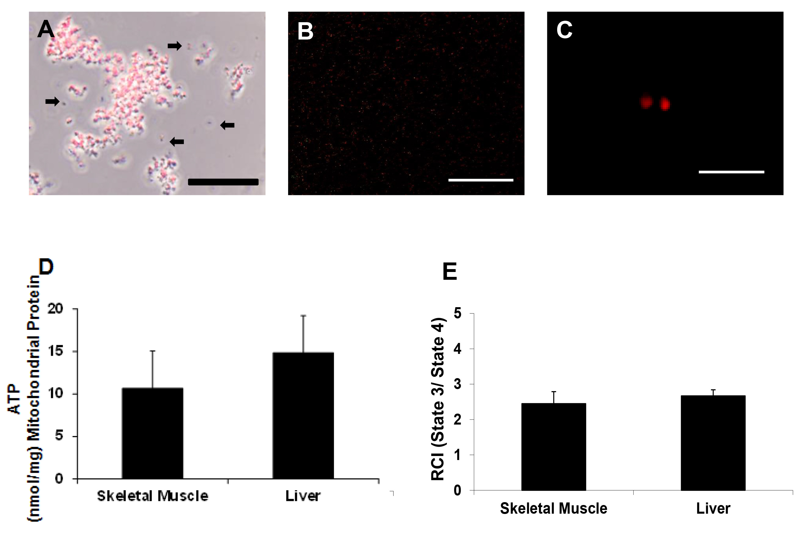
Following on from my post about the problematic field of mitochondrial transplantation, another paper was bought to my attention, in which the authors injected mitochondria into the eyes of human patients. The paper is posted as a pre-print on Research Square, which is Nature Publishing Group’s pre-print platform for papers under review or in process at their journals.
Dodgy billionaire funded pseudo-science aside, many of the problems I already outlined about mito’ transplant are present in this new paper, including an apparent claim that mito’s were counted using a hemocytometer (not possible). The mitochondria were isolated using the highly problematic serial-filtration method – the one that makes a kind of mito-ish soup with lots of blood contamination plus some residual BSA and bacterial protease for good measure.
Notably, the results section (page 7) of the paper claims that the yield of mitochondria was as follows…
“Hemocytometer counts gave 7.5 x 10^7 mitochondria for the first product, administered to the right eye, and 2.5 x 10^7 for the second, administered to the left”
So, the average total yield was ~5 x 10^7 mito’s (average of 7.5 and 2.5). Suspending all of these mito’s in 100 microliters (0.1ml) of buffer would result in a working concentration of 5 x 10^8 per ml. Later on in the methods section (page 18), we learn that…
“The final pellet was resuspended in approximately 100 microliters of cold Solution A to a working concentration of approximately 2 x 10^9 mitochondria per mL”
How did the effective concentration of mitochondria magically go up by 4 fold (i.e., 2×10^9 / 5×10^8)? Could it possibly be that hemocytometers cannot be relied on to count mitochondria, and in reality the investigators don’t really know how many mito’s they’re injecting?
Other problems surround the volumes used for testing vs. administration to the patient. It appears only 50 microliters were injected (using a 1ml syringe, which a silly way to do this and carries a risk of air bubbles), leaving only 50 microliters for all the various sterility and mito’ function tests that were needed. That’s odd because…
“Rapid microbial detection was by STAT Gram stain on an approximately 100 microliter retention aliquot”
How did they suspend the mito’s in 100uL, inject 50uL to the patient, AND have 100uL left over for microbial testing, plus endotoxin testing, MitoTracker Orange staining, and compendial sterility testing?
Given the known impact of antibiotics on mito’ function (including fluoroquinolines), it’s also odd that they chose to treat the patient with such an agent (moxifloxacin) beforehand. The eye surface was also treated with iodine (kills everything), plus neomycin/polymyxin/dexamethasone ointment. Neomycin is toxic to mitochondria. So is polymyxin. Vancomycin was also applied (you guessed it), along with ceftazidime and voriconazole. It’s a wonder there were any mito’s left after encountering this soup of antimicrobials.
Track Record
The first author of the new paper was also lead author on a previous clinical trial for mitochondrial transplant in acute cerebral ischemia. Naturally, that study prepared mitochondria using a “validated protocol” – the serial filtration method in the 2014 JoVE paper.
Notably in the ischemia study they measured mitochondrial viability using a Resazurin Cell Viability Assay Kit (from Biotium). This is nothing more than a simple NAD(P)H oxidase kit, essentially the same as an MTT assay. Apparently the kit was used “following the manufacturer’s instructions”, which is odd because the manufacturer doesn’t say anything about using it for isolated mitochondria instead of cells.
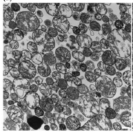
Of course, the paper shows a nice picture of some mito’s under electron microscopy…
…and then proceeds to claim “minimal contamination from non-mitochondrial components”, on the basis of absolutely no quantitative measure whatsoever.
The other mito’ data are similarly crap. There’s an N=1 for TMRM staining. As already discussed they measured ATP in the prep, finding a concentration of 3 pmols per microgram. This apparently “aligns with expected viability benchmarks for isolated mitochondria” with a reference to this paper. The values reported therein are 11 for brain mito’s and 0.15 for liver mito’s, but there are no data for muscle mitochondria.
Somehow, “our muscle mito’s had some random amount of ATP that’s somewhere in the middle of what people found 25 years ago in mito’s from completely different tissues” doesn’t have the same ring of authority to it.
I think the only sane response to this latest example of mito’ transplant insanity is that I would rather stick pencils in my eyes!
(Warning – this post contains swearing. Once you’ve read it you’ll understand why)
Anyone who follows mitochondrial research closely will be aware of a phenomenon that’s appeared in the mito’ literature in recent years – namely the concept that mitochondria can be isolated from a tissue or cells, then delivered to a tissue or intravenously to combat all manner of disease conditions.
Yes REALLY!
Such autologous mitochondrial transplantation shares many parallels with the concept of stem cell transplantation, including (as will be discussed here) a propensity for crap methods and data.
The first studies suggesting this strategy came from the McCully lab at Harvard’s Boston Children’s Hospital, showing that injected mitochondria could lower infarct size in a rabbit model of myocardial ischemia (heart attack) by around a 50%. There has since been a plethora of studies showing benefits of mitochondrial transplant for ischemia in other tissues (e.g., brain, liver) plus to treat many other conditions. Here’s a review covering various studies so far in the field of ischemia. The mechanistic claims behind this approach are quite bold – namely that mitochondria can be taken up into cells and can improve mito’ function within them.
Despite some promising findings, there has also been a steady backlash, questioning the fundamental mechanism by which any of this could work. Some of the specific issues raised are:
(1) Mitochondria are known to regulate their Ca2+ levels within a tight window, and elevated Ca2+ (~100uM) is toxic to mitochondria, triggering the permeability transition pore, a key step in cell death. The free Ca2+ concentration outside cells (~2 mM) makes it highly unlikely any mitochondria could survive in such an environment, before entering cells.
(2) The amounts of mitochondria that can achieve these effects are minuscule. The AJP paper mentioned above delivered ~6 million mitochondria to the infarct region of a rabbit heart. If one mito’ is a 0.3 micron diameter sphere (to use value determined by the same authors) then it has a volume of ~1.4 E-20 m^3 (14 attoliters). A typical cardiomyocyte (10x10x100 microns) has a volume of 1 E-14 m^3 (10 femtoliters), and cardiomyocytes are ~30% mitochondria by volume. So, a heart cell would contain about 200,000 mitochondria, and therefore 6 million mito’s is about 30 cells’ worth. Even if we pull a commonly cited value from the literature (based on zero evidence) that a heart cell contains 8000 mitochondria, then 6 million mito’s is still only 750 cells’ worth. That’s not much.
(3) Due to the evolutionary connection between mitochondria and bacteria, mitochondrial proteins are highly immunogenic, and indeed many auto-immune diseases such as lupus exhibit autoantibodies against mito’ proteins. This has led to proposals that injected mito’s may simply trigger an immune response that accounts for their effects, as discussed extensively here. Rigorous experiments in immune compromised mice to rule out this mechanism have yet not been performed.
In sum, it’s fair to say that a non-zero number of people in the mitochondrial research field are highly skeptical about this whole mito’ transplant thing, especially when it comes to the details of how it actually works.
Onward Into Humans!
Despite these concerns, the first human clinical studies of mito’ transplantation (MT) have already occurred in pediatric patients. The subjects of this 2021 paper were pediatric patients given ECMO (extracorporeal membrane oxygenation) to treat cardiac ischemic injury that occurred during a surgical procedure, with the ischemia itself treated by revascularization, and MT performed 1 day later. Some patients in this paper were part of an earlier published study.
The enrollment period for the study is given as 2002-2018, and the paper states that MT between May 2015 and July 2016 was under an IRB protocol, and then between July 2016 and December 2018 patients were enrolled in a clinical trial. The NCT record lists a trial start date of August 2017, so there’s a period of over a year (July 2016 – August 2017) in which patients were apparently given MT while enrolled in a clinical trial that hadn’t actually started yet.
A somewhat bigger problem with these time frames is that the 10 MT patients must have been during the years 2015-2018, whereas the 14 controls were spread across a 16 year period (2002-2018). This “era effect” (which, to be fair, is acknowledged in the paper) raises the possibility that at least some of the effects of MT are attributable to changes in the clinical management of these patients since 2002.
Nevertheless, additional human clinical trials are underway in stroke patients, and on we march. Here’s a study taking mito’s from urine stem cells and injecting them into oocytes for IVF (taking the piss?). This one does the same but gets the mito’s from adipocytes instead. This one takes mito’s from umbilical stem cells and uses them to treat polymyositis (a rare muscle disease). It ended in 2024 but hasn’t posted any results yet. Another study uses mito’s from placenta to treat the mito’ disease Pearson Syndrom. It’s run by a company called Minovia therapeutics, who apparently are “Harnessing the therapeutic power of mitochondria.” Here’s another trial of mito’ transplant for Parkinson’s disease, and another one for patients undergoing cardiac bypass graft surgery.
Naturally, there are lots of biotech’ companies in this area, because there’s money to be made in selling people on a cool new therapy idea. In addition to Minovia just mentioned, we have Cellvie, Mitrix Bio, Paean Bio, LUCA sciences, MitoSense, and TaiMito. It is somewhat troubling that many of these companies have well-known and highly respected members of the mitochondrial research community on their scientific advisory boards.
Overall it’s fair to say that the cat is well and truly out of the effing bag when it comes to shoving mito’s into humans! Surely the underlying animal data to support such trials is solid, right?
Extraordinary Claims Require Extraordinary Evidence, But Unfortunately The Evidence is a Bit Shit.
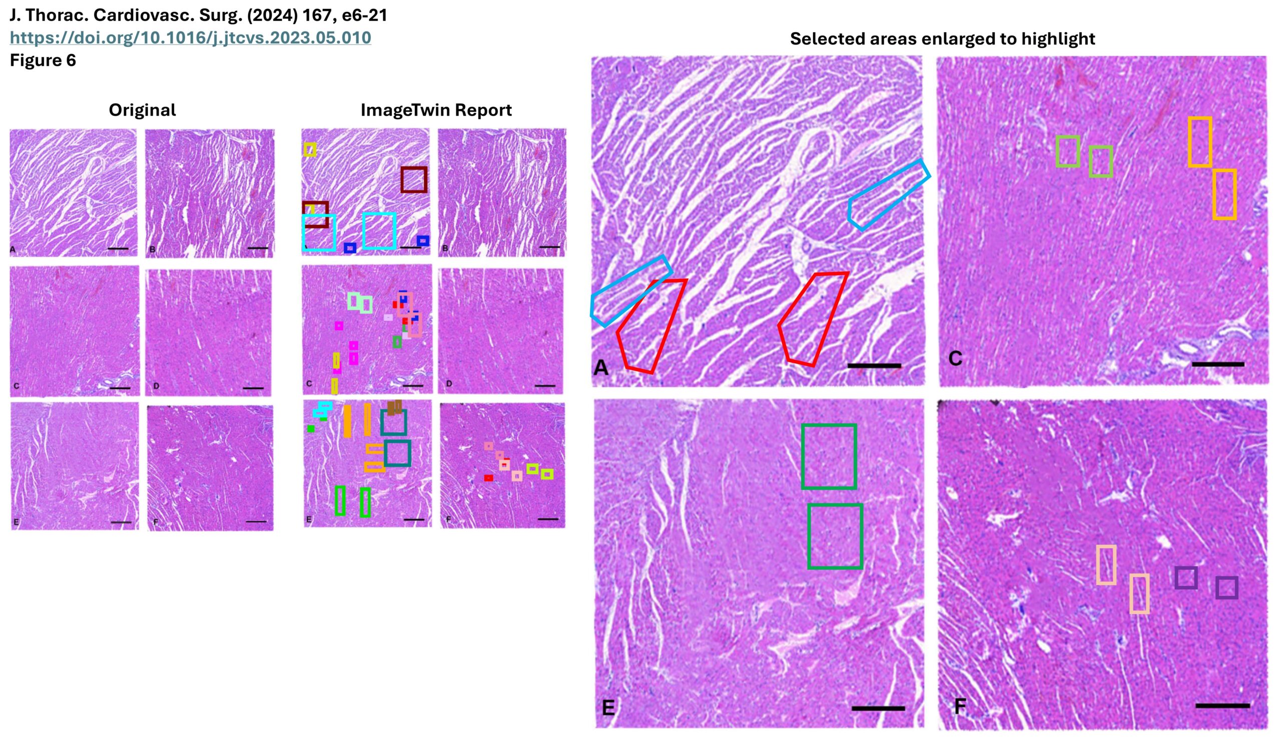
Here’s a study looking at mito’ transplant in pigs from 2023 (i.e., after the human studies mentioned above). As seen below (and explained in more detail on PubPeer) someone seems to have a passion for using photoshop when preparing H&E stained microscopy images…
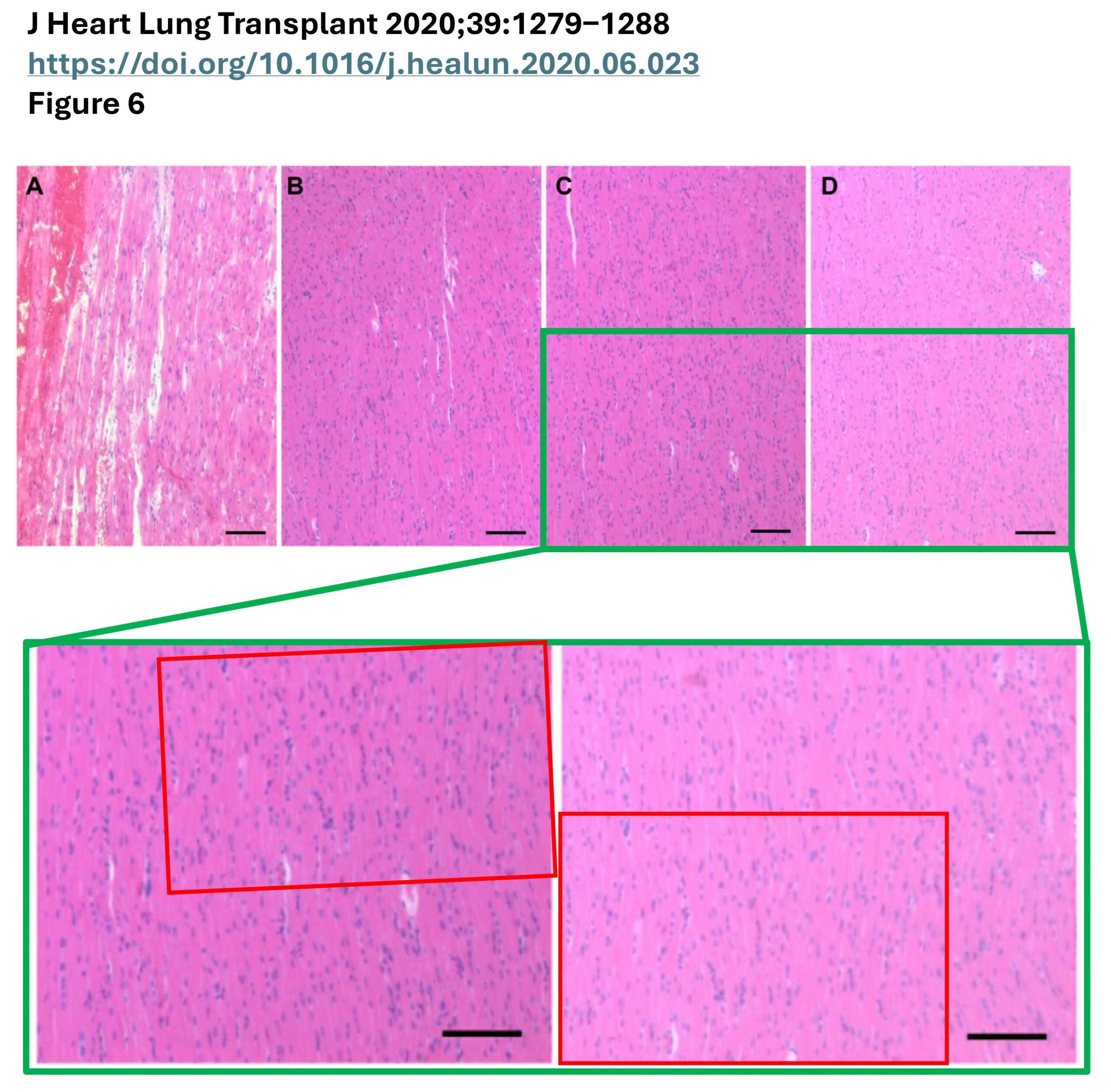
Similar shenanigans are seen in another 2020 pig paper from the same group…
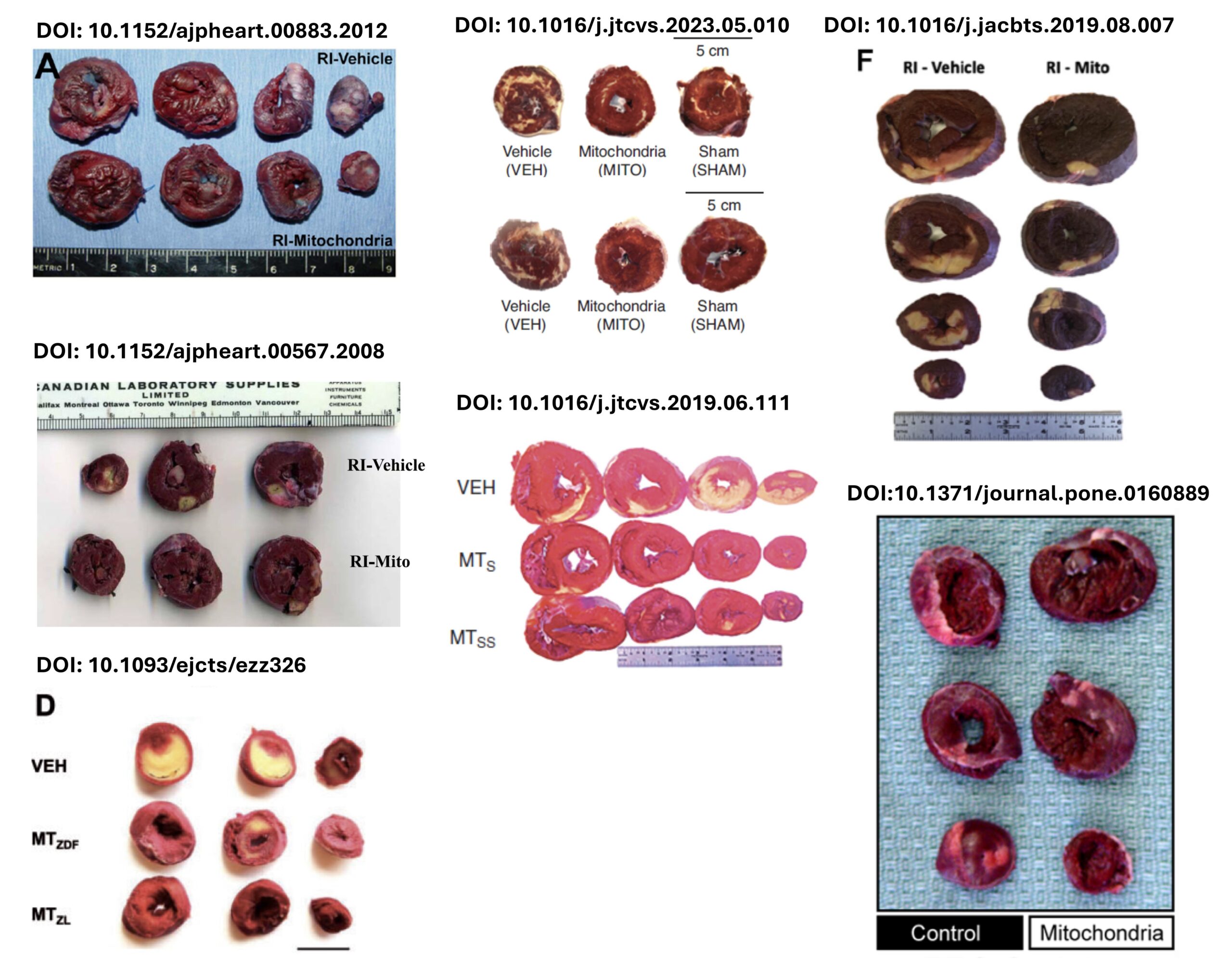
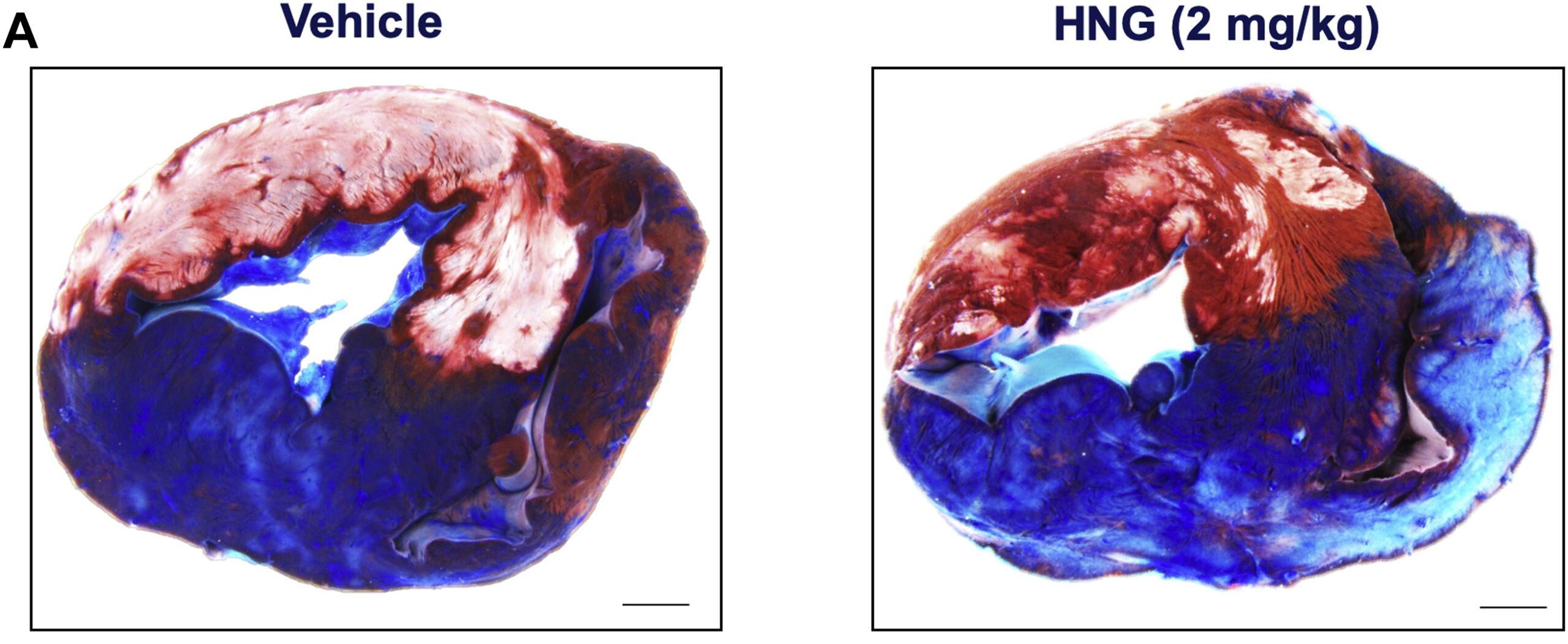
Aside from evidence of data manipulation, there are basic concerns about the quality of the other data. For example, here are a selection of images of cardiac infarcts from mito’ transplant studies across several labs and animal models…
The images are poor quality and out of focus. Some of them use inches for measurement (what is this, the darkages?), some are washed out, others have weird shadow lines suggesting they were not photographed together. In some the hearts are squashed or the ventricle lumen is filled with clotted blood. Some have white reflections indicating they were photographed in too strong a light (which gives a false positive signal when quantifying infarct area). The ones on the top left look like they were sliced with a hacksaw. These are pig, rabbit and rat hearts, so they’re all large enough to photograph at high resolution. There are often no details provided on how infarct size was calculated from these images.
For comparison, here’s what a high quality pig infarct image looks like, from a paper by David Lefer’s lab. You can clearly see the contrast and resolution, and the cardiac anatomy (left and right ventricle).
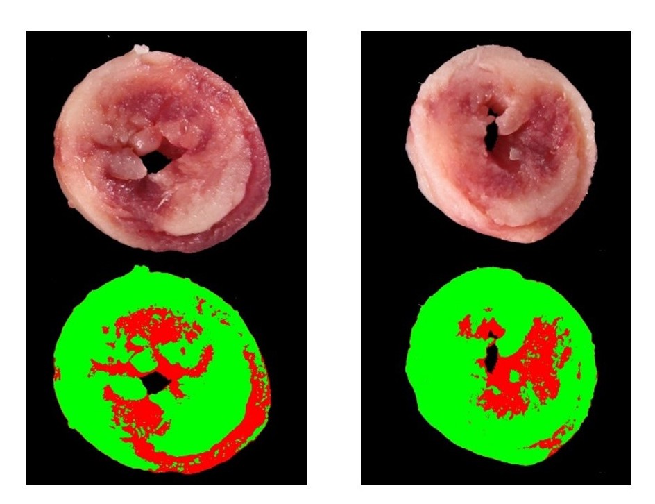
Here are some infarct images from much smaller mouse hearts in my lab (from here). You can see the contrast between white infarct and red live tissue. You can see the lumen of the LV, and the RV toward the lower right. Each image is accompanied by a pseudo-color map showing how pixel counts were used to distinguish infarct vs. live tissue.
So, it’s reasonable to say that the primary animal data supporting the notion of putting mitochondria into human hearts is of questionable quality. There are a lot more examples of problematic data further down the page, but first…
Let’s Make Some Mito’s!
Consider the methodology used to isolate mitochondria for transplant. Most studies in this area use a method published in 2014. A key step in the protocol is the use of a protease to digest muscle tissue. This technique was pioneered by Charles Hoppel in the 1970s and is routinely used in mitochondrial research because it releases mitochondria trapped between the myofibrils in muscle cells. The protease of choice is Subtilisin A, obtained from Bacillus licheniformis (in reality from Sigma). Fun-fact… a version of the same enzyme is a component of modern laundry detergents.
One of the known problems with using proteases to isolate mitochondria is it’s almost impossible to get rid of the protease in subsequent wash steps. A common solution (used here) is to dump in a large quantity of another protein such as bovine-serum-albumin (BSA) to give the protease something else to chew on so it doesn’t keep on digesting and destroy the mitochondria. One would hope that any proteins used during the mito’ prep would at least be manufactured to clinical (GMP) specifications. Alas, the BSA used here is simply heat-fractionated cheap stuff from Sigma, which is loaded with lipids and various heme-related pigments. Even though Bacillus licheniformis is gram-positive and doesn’t generate endotoxin, I wonder if the patients undergoing this procedure are told that the “autologous mitochondria” being infused are very likely contaminated with remnants of a bacterial enzyme plus a crude fraction from cow blood?
After digesting the tissue, many mitochondrial preparations use differential centrifugation to separate mitochondria from other cell components. First a low speed spin (~1000 x g) gets rid of cell debris and nuclei, then a series of high-speed spins (~10,000 x g) pellet the mitochondria and wash away cell components of a similar size that would contaminate the prep’ (fragments of ER, SR, peroxisomes, microsomes, etc).
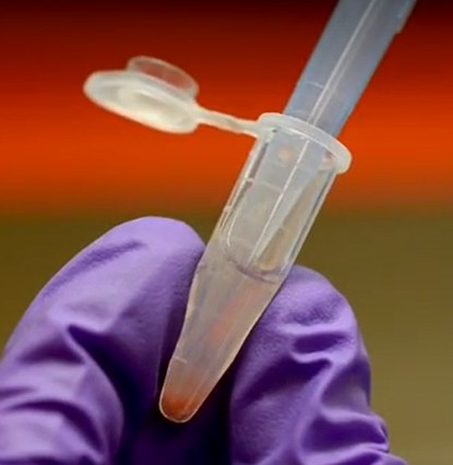
Instead, this method simply passes the cell homogenate through 3 filters (2 x 40 micron, then 10 micron) then a centrifugation step to pellet the mito’s. This is problematic because filtration will get rid of things bigger than the filter size, but anything smaller will pass through – including all those mito-sized membrane fragments I just mentioned, plus the bacterial protease and BSA. And don’t forget the blood! Yes lots of blood, as evidenced by the red color of the mito pellets in the accompanying video. Here’s a screen shot – that red stuff at the bottom of the tube is blood. Mitochondria are light brownish-green, not red.
There’s also a mis-match in the paper’s claims about particle size and purity of the mito’ preparation. It is claimed 0.18g of tissue yields 2 x 10^10 mitochondria, and the mito’s are 0.3 microns across. That’s a total mito’ volume of just 0.3 microliters, which should not be a visible pellet, and yet the video clearly shows a pellet (maybe 50 microliters) yielding a milky suspension. The paper makes bold claims about the purity of the preparation, but includes no measurements to show the removal of other contaminating cell fractions (e.g., western blot for other membranes). There are some electron micrographs, but no description of how such images were analyzed to arrive at the claimed “<0.001% contamination by non-mitochondrial particles.” A simpler interpretation is that the mito’ prep cannot possibly be 99.999% mitochondria, and it is very likely contaminated with other membrane fractions and blood (for the record, red blood cells can easily pass through a 10 micron filter).
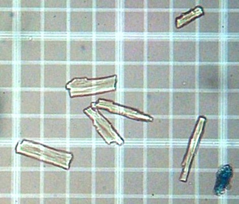
The method paper also claims mitochondria were counted using a hemocytometer. Here’s a picture from my lab of some cardiomyocytes on a hemocytometer grid. Each small square (bounded by single lines) is 50 microns across. Those small specks of dirt are maybe 2-3 microns across. I might be almost blind, but I don’t think anyone with 20:20 vision could reasonably see and accurately count a bunch of 0.3 micron particles at such scale.
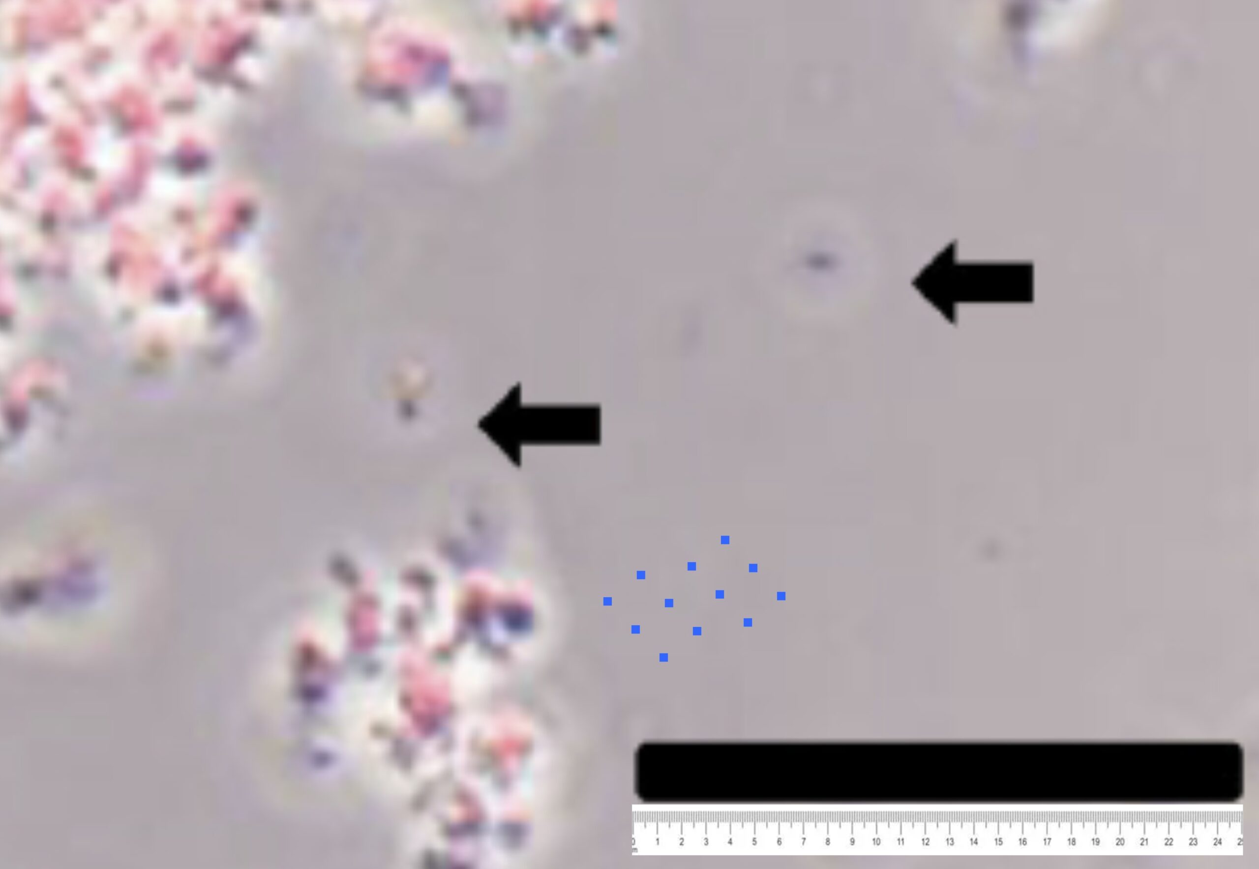
An image from the paper shows some mito’ particles accompanied by a single black 25 micron scale bar. I’ve added a ruler below the scale bar, then used it to draw in some actual 0.3 micron particles – the blue dots. Do the red things (mito’s) look like they have an average size the same as the blue things?
Lastly regarding methods, I’ll note that before this JoVE paper, the 2013 AJP mito transplant paper isolated mitochondria from skeletal muscle in the chest wall. It refers to an earlier 2009 AJP mito transplant paper for the method. That paper isolated mitochondria from heart tissue, and lists 2 references for the method. The first of those papers has no methods section and no information on how to isolate mitochondria (although it does refer to a paper that used differential centrifugation). The second is a review article about cardioprotection, with no methods. Likewise, the 2009 AJP paper cites a method to measure mito’ respiration, but the cited paper is on a different topic and says nothing about mito’ respiration.
So, it seems the animal studies used to support going to clinical trials relied on a crude mito’ isolation method from various tissues, and then a new and very different filtration method was invented and is being pushed for human studies, despite very little evidence that it actually makes anything resembling mitochondria.
But What About ATP ?!?!
Key questions for anyone preparing mitochondria include… Do they consume oxygen? Do they have a membrane potential? Are their membranes intact? Can they make ATP? This last question appears to have been mis-applied by the proponents of mito’ transplantation.
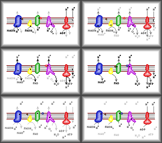
All basic biochemistry or metabolism books have a chapter on mitochondrial oxidative phosphorylation (Ox-Phos). Here’s the short version… (i) The Krebs’ cycle burns metabolic acids to generate NADH. (ii) The electron transport chain passes electrons from NADH through respiratory complexes I-IV and onto oxygen. In doing so, the complexes pump protons out of mitochondria, generating a proton gradient or membrane potential. (iii) Oxygen is the terminal electron acceptor, and gets consumed and turned into water during this process. (iv) The energy in the proton gradient is used by complex V to make ATP from ADP and phosphate. Importantly, this whole process relies on mito’ membranes being intact. Here’s an animated GIF I made when I was a grad student more than 30 years ago…
For mitochondria to be functional, what matters is the RATE at which they generate ATP. Dead or non-functioning mitochondria still contain ATP, but it’s the ability to MAKE ATP that matters. The proper way to do this (as I did a mere 27 years ago) is to incubate mitochondria with a substrate (to feed the Krebs’ cycle) plus some ADP, then take aliquots at regular time intervals, crash out the protein (to stop any ATP from being consumed) then measure the ATP. Finally, plot a graph of ATP vs. time and calculate the slope (rate). Ideally you do this +/- an inhibitor of complex V (oligomycin) to make sure the ATP is actually coming from Ox-Phos.
Is that what people isolating mito’s for transplant are doing? Nope. Instead you just slap the mito’s in a plate reader with some ATP kit reagents and a lysis buffer to break everything open, leave it for 10 minutes and do a single end-point measurement. Let me be abundantly clear – what this method measures is how much ATP the mitochondria contain. This has FUCK ALL to do with how functional they are, and the rate at which they make ATP!
The assay does not (and indeed can not) measure mito’ function, because everything is blown to shit by the lysis buffer. The fact this is now the standard method for making mito’s for transplantation (136 citations so far) is shameful. The fact it got past peer review indicates the quality of that process at JoVE. Surely human patients deserve better than this?
Why Isolate Mitochondria When You Can Grow Them?
In 2022, a paper claimed that infusion of mitochondria can improve function in the hippocampus of aged mice. Unfortunately, as documented on PubPeer, the western blots were a giant mess, which greatly undermines confidence that the authors know what they’re doing.
This is unfortunate, because the lead author on the paper Benedict Albensi, is a key scientific advisor for the company Mitrix Bio. Mitrix takes the cake when it comes to mitochondrial science fiction – they claim to be growing mitochondria in a bioreactor, and they call them “mitlets”. For anyone remotely versed in mitochondrial biology, the notion that you can “grow mitochondria” outside of cells is complete bullshit. The reality is a bit more simple… they take stem cells, grow those in a reactor, then isolate the mito’s and package them up in membranes for delivery.
This all came to prominence recently because a group of folks calling themselves “Mitonauts” published a press release claiming that the wait is over and we should all just embrace lab-grown mitochondria to combat aging!
Those of you who know me will perhaps recall an article I wrote several years ago about the giant shit-show that is longevity biotechnology, and this appears to be nothing different. A bunch of rich libertarians arguing for right-to-try and lax regulations, so they can apply a therapy with not very good supporting data (see above), to overcome a poorly understood pathologic state. They’re testing it on themselves, which I guess is slightly better than trying it on children. Stem cells didn’t work, but surely mitochondria isolated from stem cells will work. Hey it’s your money, who am I to tell you how to waste it?
Why Inject Mito’s When You Can Just Eat Them?
Why go to all the bother of injecting or infusing mitochondria, when you can just EAT them instead? This is especially true if they’re plant mitochondria, because we all know that plant based diets are more healthy. Of course, the paper was absolutely loaded with manipulated images, as documented on PubPeer. Leo Schneider has a nice write-up of similar shenanigans from the same lab going back several years.
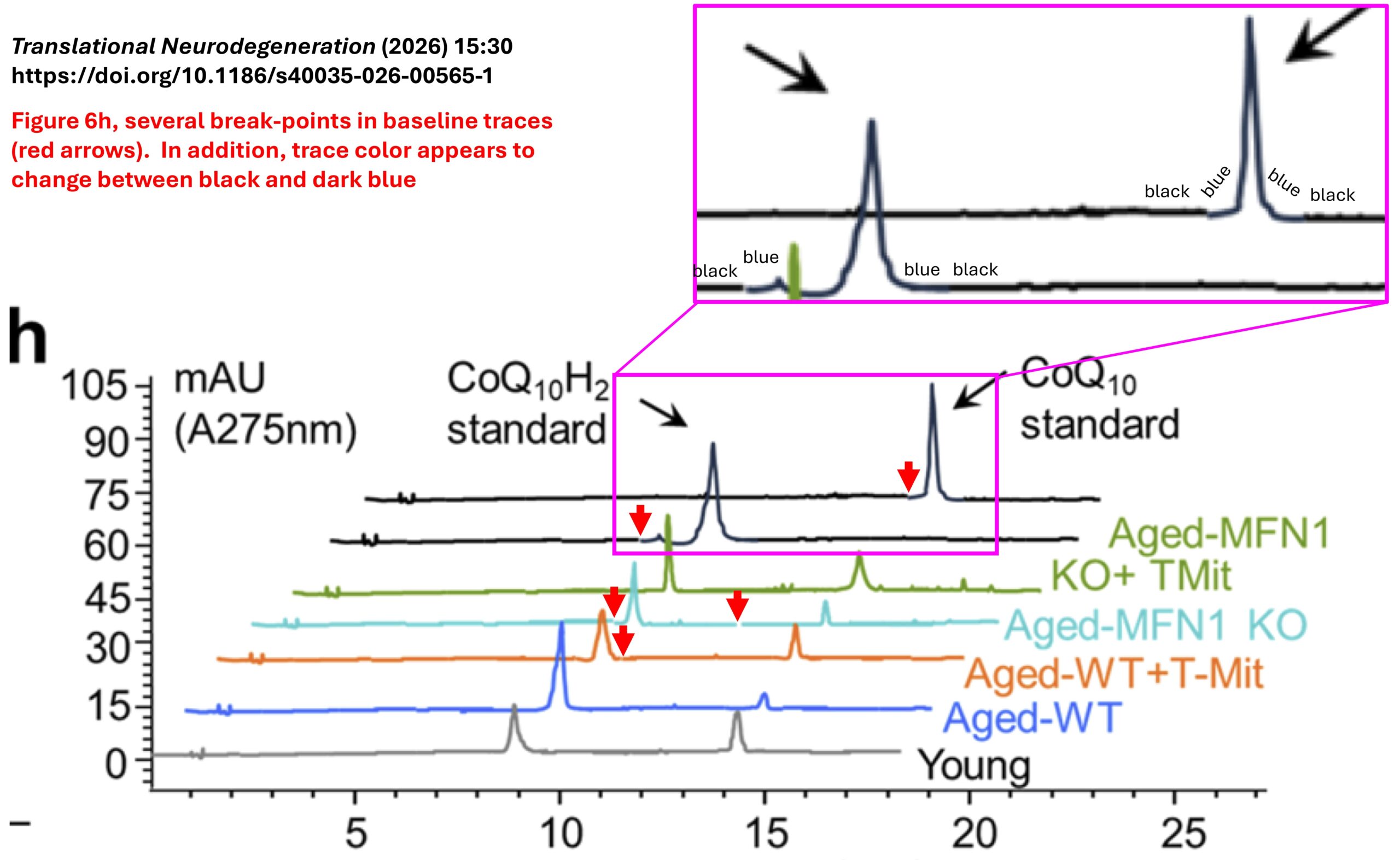
My favorite is this example below, in which the HPLC data appear to be faked by pasting in the peaks onto the baseline. The problem is whoever did this forgot to change the colors, so they ended up with a black baseline which magically switches to a blue line for the peaks, then back to black again. Both the authors and the editors of the journal were notified, and neither have responded to indicate they’re even remotely interested in dealing with this.
This Shit is Everywhere!
Lest anyone think the problems in this field are limited to a few labs and obscure journals, here are just a few more of the papers on mitochondrial transplantation in which I and others have found “problems”…
Here’s one I found this week, on mito transplant for muscle wasting. Image overlap reported on PubPeer, authors notified, and apparently this will be corrected.
This paper is about mito’ transplant to treat fatty liver disease, but (as explained on PubPeer) the authors reused some images in another paper, which doesn’t instill confidence in their lab’s data management.
Here’s another one all about mito’ transplant in melanoma, with dodgy western blots. Supposedly the authors sent a correction to the journal in February 2024, but it hasn’t been corrected yet.
Here’s a paper where they did mito’ transplant and claimed to do CPR on rats, at a rate of 300 beats per minute. That’s 5 times a second! The best gamers in the world can maybe do 6 mouse clicks a second, so the notion that anyone could do CPR with any control over depth or accuracy of compressions at a rate of 5Hz, is not credible. No response on PubPeer yet.
This paper claims tunnelling nanotubes can transport mitochondria between cells in the hipoocampus. The western blots are laughable.
This paper on mito’ transfer in the tumor microenvironment got called out for some numerical irregularities (aka. creative use of copy/paste in Excel), and then swiftly corrected. Nothing to see here, move along please.
This one is about mito’ transfer in cancer stem cells. You guess it, crap western blots again! Another paper in the same area had some problems addressed in an erratum, then other problems raised later on PubPeer which have still not been resolved. Yet another paper in the same area is very problematic.
Here’s one I found just yesterday on mito’ transplant in stroke, where the authors apparently had electron microscopy data on 94 specimens from 3 batches of mitochondria, but somehow chose to show 3 parts of the same image cropped differently as evidence.
Naturally, the field is awash with review articles, editorials and puff-pieces, talking up the whole idea of mito’ transplant and how wonderful everything is. How do you think I found most of the problematic articles I flagged on PubPeer, for this blog post? That’s right – they’re the very same one being cited to support this madness.
Summary
It’s not looking good. The entire field of mitochondrial transfer and transplantation is flooded with crap data and methods. The data from animal models being used to support human clinical trials is simply not fit for the task, and the clinical trials themselves are not much better. Money is pouring into the biotech’ industry being built off this house-of-cards. Now the whole thing is being hijacked by a bunch of libertarians and right-to-try millionaires (“mitoNUTS“) pushing for unregulated mito’ therapy for aging and who knows what else.
The field of mitochondrial research has a noble history. Shame on any bona-fide mitochondrial biologist who goes along with this crap without speaking out!
Anyone following developments at NIH will be familiar with a new category for disposition of grant proposals following initial review: CND, competitive not discussed. (Flashback for GenXers like me: not to be confused with CND, the Campaign for Nuclear Disarmament, whence the original peace symbol)
Previously, for most NIH grant review panels, prior to the review meeting applications were placed in two bins based on preliminary scores, with the top half being discussed, and the lower half being not-discussed (ND). As such, when you submitted a grant there was at least a 50% chance it would be discussed in person by a team of 25-30 reviewers (vs. only being seen by 3 assigned reviewers).
Due to the glut of proposals that were not reviewed during the federal government shutdown in fall 2025, part of the emergency measures to relieve the backlog included placing proposals into 3 bins… top 3rd discussed, bottom third ND, and now the middle third CND.
The CND designation is supposed to take account of more freedom in funding decision making within NIH, such that applications assigned CND could technically still be funded. However, I am not aware (and don’t know anyone else who is aware) of any CND application so far being pulled up for funding. Rather, the net result is that now only 1/3 of proposals get fully discussed. And there are numerous tales of grants getting discussed and getting good scores, but the funding simply not coming through, so it seems even getting a fundable score is no longer a guarantee that money will be forthcoming.
So why am I talking about this now?
As luck would have it, the competing renewal application for my long-running R01 grant was submitted in November last year and, because of the shutdown, its review was delayed by around 6 weeks from mid February until late March. It ended up in the CND bin, which means we now have to resubmit for the July NIH deadline. Among the review critiques, one reviewer stated that productivity in the prior grant period was strong, while another claimed it was weak – for the record I published 24 papers during this time, when this was my only grant as a PI. The same reviewer listed “studies are mainly mechanistic” as a weakness. Also our hypothesis being “mostly based on literature or preliminary data” was seen a weakness.
So here we are in mid 2026, with my current (and only) grant in no-cost-extension, running out of funds rapidly. The lab is running on fumes, with only me and a grad student, no technician, no post-doc or other personnel. A 23 year track record of NIH funding about to come grinding to a halt, with no idea how we will keep the lights on a few months from now.
The silver lining is that since last November we’ve obtained some really cool new data on circadian responses to heart attack that I hope will assuage some concerns of the reviewers. We just have to hope that the resubmission will be properly read, so the message can get through that my lab can still add value to the field.
I just posted a new pre-print reporting on a small investigation in the area of image manipulation and research misconduct. It centers on content from the Journal of Cancer, which is published by the Australian open access publisher Ivyspring.
The TL/DR version is as follows: If your journal does a poor job of screening for garbage pre-publication, it doesn’t really make a difference whether you charge a fee for authors to clean up their own mess.
Charging what now??
One of the notable things about this journal (and others from the same publisher) is that until recently they charged an additional fee for the publication of a correction/erratum to an already published article. Such fees are generally frowned upon by the Committee on Publication Ethics (COPE).
Not only could such fees discourage authors from coming forward to correct any problems identified post-publication, it could be hypothesized that they actually create a disincentive for the journal to address problems identified during pre-publication. After all, if you just say nothing and the problem is subsequently found, that’s more sweet revenue for the taking ($AU 1,750 or about $US 1,200 at the time of writing).
In late 2024 the journal abandoned this fee policy, which provides for a convenient way to test a hypothesis. Simply look at the rates of problematic material before and after the policy change, to see if it made any difference to the incidence of problems that need correcting.
ImageTwin-AI to the rescue
Of course, it’s no small undertaking to manually screen hundreds or thousands of papers for problem images. Luckily these days we have AI-assistance tools such as ImageTwin-AI to help with that. There are several such platforms available (including some that are problematic), but ImageTwin is among the best I’ve encountered and has an easy-to-use platform. Previous studies have demonstrated the utility of these platforms in the right hands. FULL DISCLOSURE – I was an early tester for ImageTwin, and so have access to it as a courtesy from the site owners (thanks Markus!)
What was done?
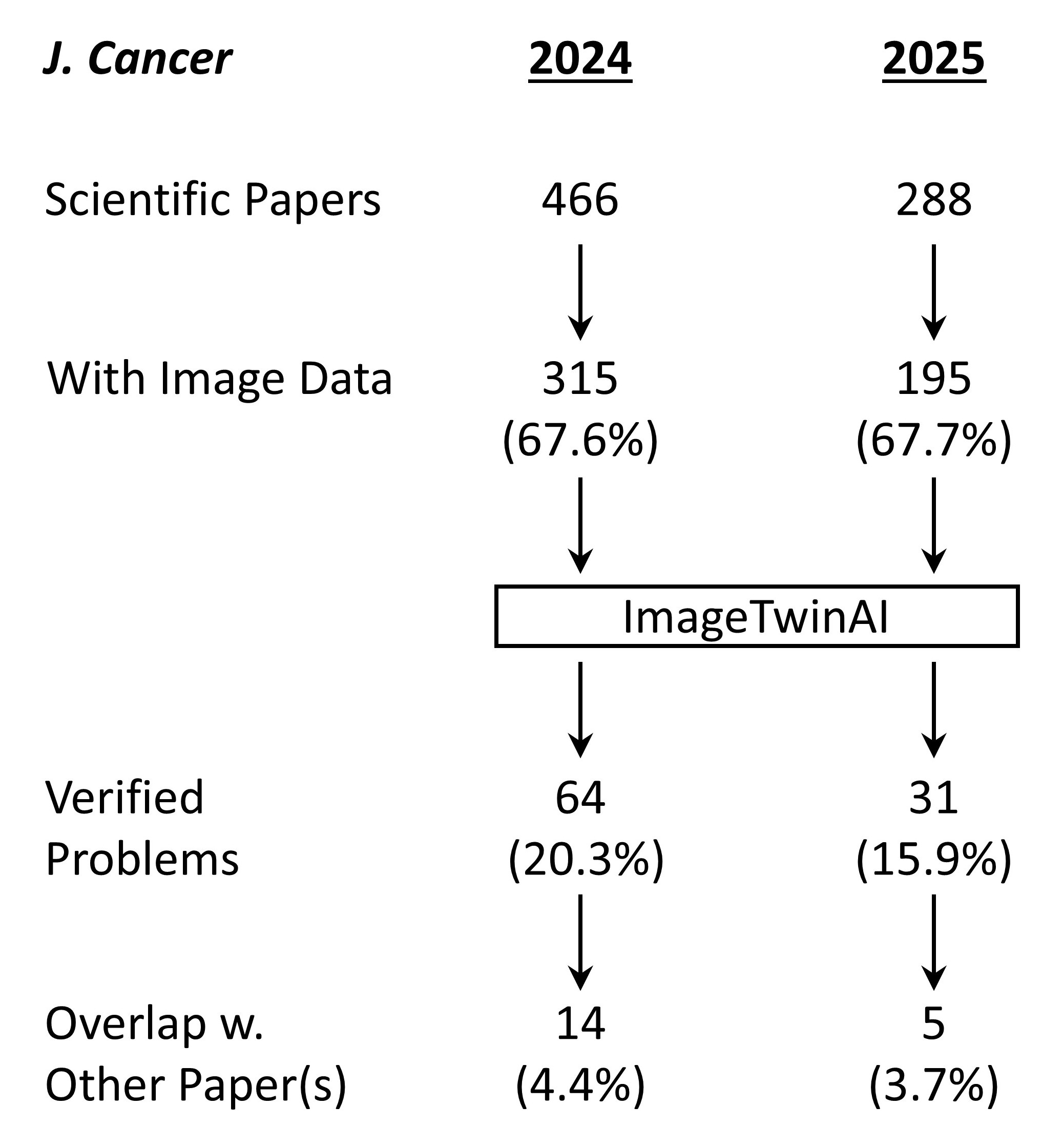
Shown above is the workflow for the study. A total of 754 papers were of the type suitable for screening – meaning they were original scientific papers rather than review articles, editorials, consensus statements or other journal content. Of these, 510 contained image data and were fed into ImageTwin (usually in batches of 4-6 at a time). Every “hit” was manually verified, then annotated in powerpoint to highlight the problem(s).
A number of problem images were flagged by ImageTwin but removed from the analysis. This included the use of IHC and other images from publicly available databases such as the Human Protein Atlas (HPA). Likewise, using the same image to represent the same experimental condition (such as control cells) within a paper cannot really be construed as a huge problem. It’s not best practices, but it’s also likely not misconduct (the exception here is when a single western blot loading control image is used for dozens of blots, which is simply physically impossible).
What was found?
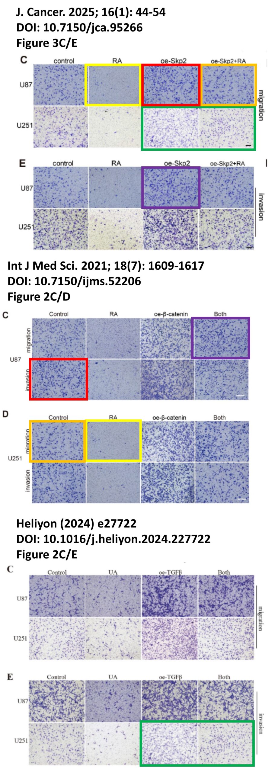
The platform found 95 papers with evidence of inappropriate image manipulation. This included 19 papers with overlapping content from completely unrelated journals or papers. This latter group really is the gold value proposition of ImageTwin – it would have been virtually impossible to find such image matches manually. Here are a couple of examples:
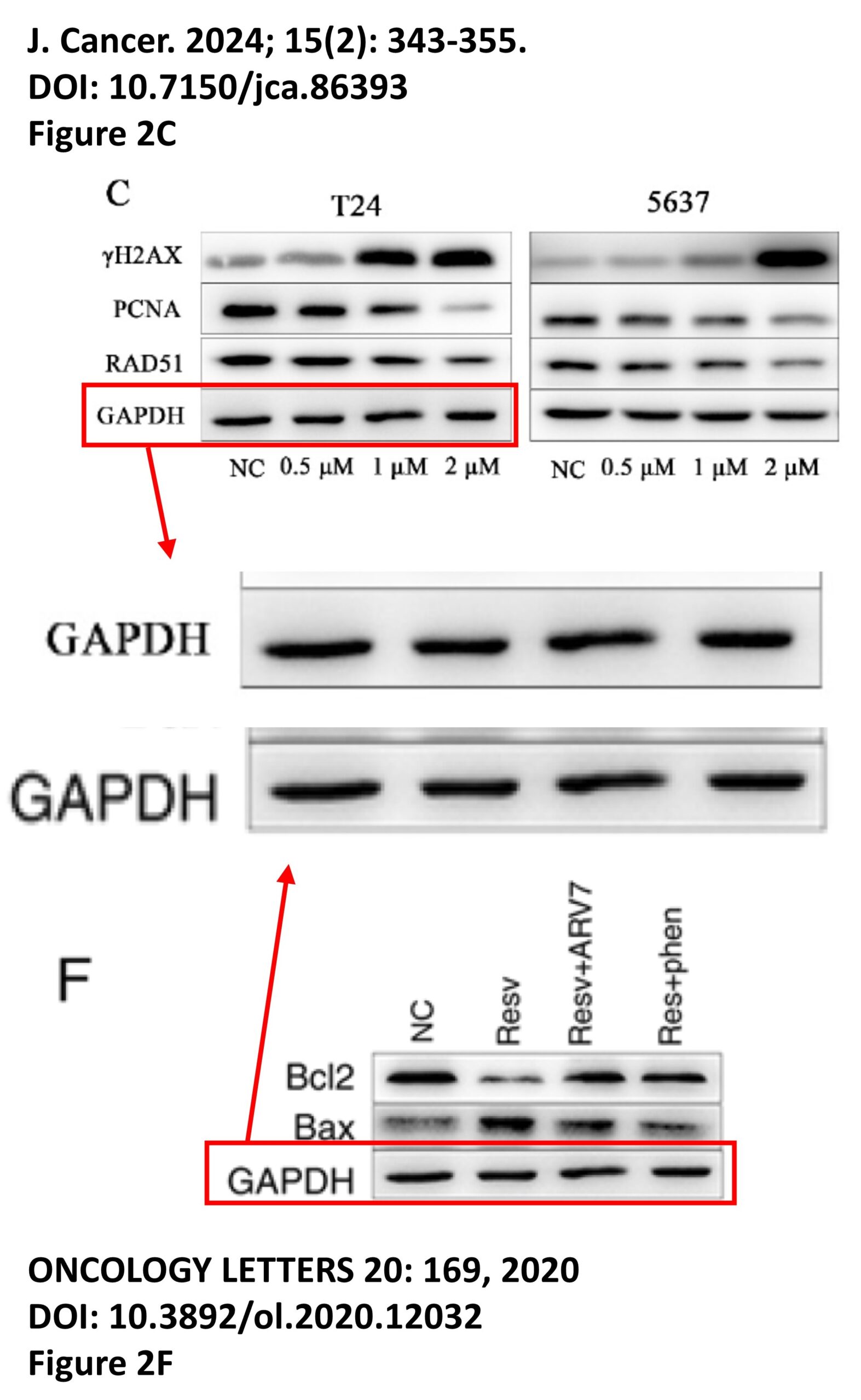
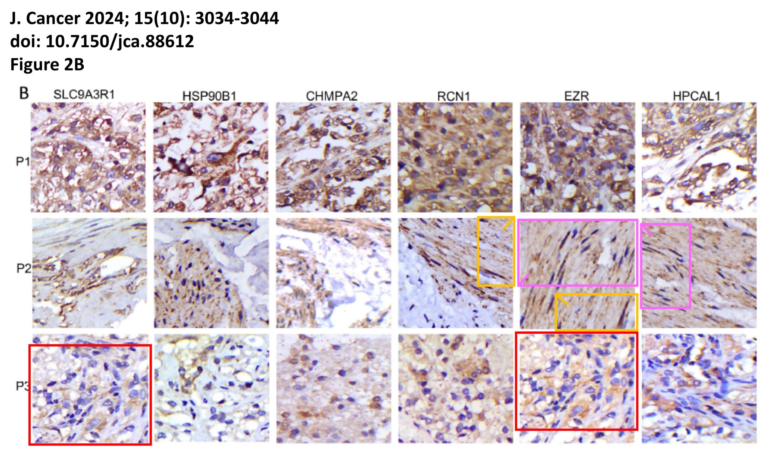
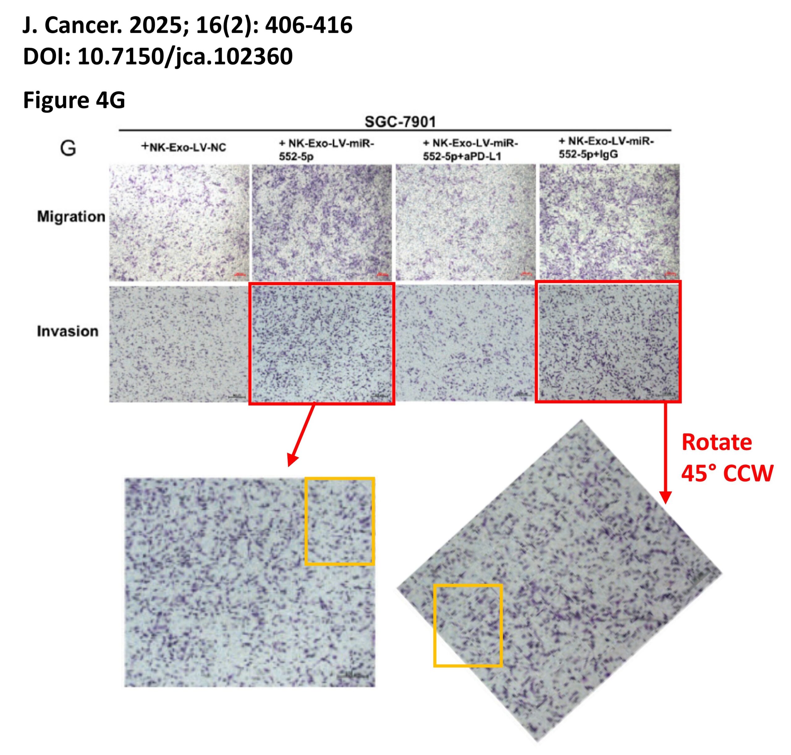
The platform also identified a host of problems within individual papers, such as these…
For those interested, the complete documentation of all problems is available on FigShare, comprising 2 PDFs with the annotated images, plus an Excel spreadsheet detailing the 153 separate examples of image manipulation.
What about the original question?
Overall the fraction of papers with problematic images fell from 20.3% in 2024 (while the correction fee was in place) to 15.9% in 2025 (after the fee policy was rescinded), suggesting only a modest impact on editorial processes at the journal.
Caveats
Of course, there are many caveats to this study, such as the fact it’s from a single journal and only a few hundred papers. It would be interesting to repeat the analysis with other journals from the same publisher, since they all had the same fee-for-correction policy in place until recently.
Another caveat is that of course this data set doesn’t reveal anything that goes on at the editorial level. Who knows why or when the publisher decided to stop charging for corrections? It is notable that COPE re-jigged their guidelines on fees in late 2024, but that may be pure coincidence. Personally, I would consider it highly unlikely that on Jan 1st 2025 the editors woke up and said “hey we’re not charging for corrections any more, so get out there and start flagging more stuff during peer review.” If they did, it wasn’t very effective.
One final caveat is the different sized cohorts of papers, with the 2025 volume of the journal publishing ~38% less content than in 2024. This could have been due to simple external factors such as fewer submissions, as scientific funding around the globe becomes harder to obtain. Or it could be because the journal raised their APC to $AU 4,000. Either way, 16% problem papers is still a big number, and not a lot to be proud about.
What’s Next?
As time permits, I may get around to manually posting each of these items on PubPeer, which is unfortunately not yet an automated process. As with all AI tools, the “Trust but Verify” mantra is critical, hence everything in the paper was manually verified and annotated prior to publication. That’s really #1 on my wish-list for these types of analysis, an auto pipeline to go from discovery to online PubPeer documentation in a single click. It’s hard to reconcile such a process with robust human-in-the-loop verification.
Of course if anyone at the journal is reading this, feel free to pull all the original data files from FigShare (it’s all neatly organized for you) and start retracting the worst examples. And maybe consider a subscription to ImageTwin?
There’s also the “cost” to consider. Not just the monetary cost of screening large numbers of papers, but also the externalities of AI compute, including data centers, water usage, carbon footprint. Plus all the other problems of the genAI industry at large, as explored at length by one of my favorite bloggers, Ed Zitron. Whether any of these platforms will be profitable before they run out of venture capital remains to be seen, but personally I’d rather see AI being used for applications such as this, instead of generating pictures of a cat dressed as Lady Gaga playing the flute while riding unicycle.
Summary
What this study really highlights is the great utility of platforms like ImageTwin-AI for rapidly screening papers and discovering image problems. It would have taken forever to do this manually.
The prevalence of problem images in this cohort of papers (18.6% overall) was similar to a previous study from Sholto David on content from a different journal (which found 16%), and agrees with the contention from James Heathers that “1 in 7 scientific papers are fake.” In particular, the 4% of papers that contained images duplicated from unrelated journals are likely candidates for Paper Mill papers, and warrant further investigation on that front. Hopefully they get retracted ASAP.
Lastly, this study really doesn’t support any kind of causative link between following COPE policies and actually driving out problems from a journal. It takes more than simply signing up to an industry lobby group and paying a membership fee, to cleanse the literature of garbage.
This story spans several years between 2010 and 2014. It was sitting in my drafts folder for over a decade, so some links may be dead and the current status of various people or companies or science may no longer be accurate.
Here’s the short version…
My lab got a drug from a company via a material transfer agreement (MTA)
We found some really cool results
We tried to publish results
The company tried to sue us
The company went bankrupt
We published the paper anyway
Rival paper from former company employees came out, doesn’t cite us
Background on Conflict of Interest in Basic Science
For life scientists in academia, one of the most common examples of potential conflict-of-interest (COI) is receiving resources from a drug company to do a research project. Sometimes this takes the form of money directly intended to fund the research. Other times the reward can be a position on the scientific advisory board of the company, or being granted access to rare chemicals/reagents that are not commercially available to everyone. In the US, such support is usually required to be disclosed both to the employing University, and federal or other funding agencies.
Usually when the resource being shared is a new drug, companies like to control what happens to the material, such as whether/how the academics can discuss the results with others, including publication. Such legal matters are usually dealt with via Confidentiality/Non-Disclosure Agreements (CNDAs) and Material Transfer Agreements (MTAs). These documents are drafted up jointly between the lawyers in the company and the University’s Technology Transfer Office. Herein, I discuss an example of such an agreement gone awry….
The research that led us to request an MTA for a new drug
My lab’ has a long-standing interest in mitochondrial K+ channels, and in our work we’ve often used a drug called NS1619, which is reported to specifically activate mitochondrial KCa channels. The nomenclature is a bit odd here… a family of channels is encoded by the Slo genes, of which there are 4 isotypes in mammals (Slo1, Slo2.1, Slo2.2, Slo3). The channel proteins are also called “BK” channels, since they are large conductance (big) K+ channels. They’re also often referred to as KCa channels, even though the Slo2 variants are not Ca2+ activated (they’re Na+ activated, so KNa channels). There are also some hybrid terms such as BKCa, or mBK (mitochondrial BK). Anyway, the important thing is these channels are thought to be important for protecting the heart against ischemia-reperfusion (IR) injury, and the debate mainly centers on which isotype is present in mitochondria and responsible for these effects. Other people think it’s Slo1 but our results suggest otherwise.
NS1619 was originally made by a Danish company called Neurosearch (archived link from 2019 shortly before site went dead), but is readily available from chemical suppliers such as Sigma. However, over decade or so since the drug was made available, it emerged that NS1619 might have non-specific side effects in mitochondria. So, interest in the field shifted toward a related newer compound, NS11021, reported to be more potent and specific for the mito’ channel. NS11021 is not commercially available, so in late 2011 we wrote to Neurosearch to obtain some. Following a few rounds of back-and-forth between lawyers, an MTA was executed and shortly afterward we were sent an aliquot of NS11021. The scientist within the company who liaised with us, Morten Grunnet, was collaborative and very open to discussing experimental details and results. I consider him a colleague and friend to this day, and he was a co-author on our paper that came out of this work.
Although publishing the entire MTA here would be inappropriate, below I want to highlight specifically the part describing the work to be performed in my lab using the drug. Note the mention of potential use of knockout mice for various types of K+ channel…
Appendix A: Study Protocol. The overall direction of the research project, which the study will be part of, is the identification of K+ channels in mitochondria, which are involved in cardioprotection by ischemic or anesthetic preconditioning. The aim of the study would be to investigate the role of the BK channel in anesthetic preconditioning using FVB mice. In Langendorff perfused rat hearts, the volatile anesthetic isoflurane provides protection against ischemia-reperfusion injury in a manner that is sensitive to the BK channel inhibitor paxilline. University of Rochester would like to determine if this manner of isoflurane mediated protection can be mimicked by the administration of the specific BK channel activator NS11021. University of Rochester would also like to look at the effect of NS11021 on BK channel activity in isolated mitochondria, using novel thallium-flux assay we recently published (PMID:20185796, PDF attached). None of the K+ channels in the mitochondrial membrane have been identified at the molecular genetic level. Knockout mice for various types of KCa and KATP channels may be available to us in future, and knowing which channel to look for could be greatly facilitated by the use of highly specific pharmacologic reagents.
So, we did some experiments and the data looked great. Without getting into details (all of which can be found in our published paper on this topic), the key finding was that NS11021 protected hearts against IR injury, and this effect was lost in Slo1-/- mice. Furthermore, the effect was likely mediated not by Slo1 channels inside mitochondria of cardiomyocytes, but instead by channels in intrinsic cardiac neurons.
Things get nasty
You might think if a company was touting a drug as acting on a specific target, and someone came along with data showing the effect of the drug disappears in a knockout mouse for the target, they’d be happy? Not Neurosearch, they lawyered-up and tried to stop us from publishing. They essentially said that if we attempted to publish, it would be a breach of the MTA, and they would sue for breach of contract.
Backing up for a second, during the time we were collecting our data and preparing our manuscript, we were in regular discussion with Dr. Grunnet, who was essentially our contact within the company. However, in the summer of 2012 he left Neurosearch to work for a different Danish Biotech, Lundbeck. We continued communicating while the manuscript went through several iterations, then in June 2012 we heard via Dr. Grunnet that Neurosearch were “rather reluctant to let us publish since they claim that the part including KO mice is not covered by the MTA”. Also “they have decided to perform a small study on 11021 in WT and BK KO mice with I/R in Langendorff. The primary aim is to confirm BK selectivity of 11021 and they are aiming at a small publication”. The suggested solution was that we hold off until their study was published. In other words, the company scientists didn’t want to get scooped!
Given our study was complete and ready to publish, and theirs had barely even begun, we tried explaining that it might be better to simply add the interested parties onto our paper as co-authors. In my view this was an outreach effort that went above and beyond necessary collegiality (remember – they were trying to sue us). However, we were then contacted by Søren-Peter Olesen who oversaw Neurosearch’s collaboration with the University of Copenhagen, and also holds an academic appointment there.
In the remainder of fall 2012, while our paper was being put through the wringer by journal reviewers, a conversation ensued between myself, Dr. Olesen, and the Tech’ Transfer lawyers here in Rochester. This culminated in an email from Dr. Olesen in November, stating “Neurosearch will then close the matter and conclude that you do not want any legal permission to publish on NS11021 in relation to transgenic animals”.
We had the University lawyers look over the MTA again, and they made two important conclusions… First, the language was sufficiently broad regarding knockout mice that in their view we hadn’t breached the terms. Second (and more important) the company had no right to bar us from publishing because the MTA itself made no mention of a right-of-veto on publications. The MTA simply requested we send a copy of any manuscript to Neurosearch 30 days before journal submission, for them to review and comment. We did this, and in-fact our company point-person had been kept in the loop from the earliest stages, so Neurosearch knew about these knockout mouse studies for almost a year before the paper was submitted.
Neurosearch goes kaput
Eventually, after 4 journals and 8 months of review/reject/resubmit, we got our paper published in Peer J in February 2013. But then something strange happened… Neurosearch went belly up. Well, technically the terms used were restructuring, transfer of assets, prosecution for share price manipulation. So, the company that threatened to block us from publishing no longer exists. Another company trading under the same name and with new management did emerge from the ashes of the old Neurosearch, but disappeared in 2019.
What about the company-backed study?
Olesen and colleagues finally published their version of the story in PLoS One, in collaboration with a group from the University of Tübingen. Although the new paper cites our work, there are a number of problems with it…
The paper purports to show patch-clamp data of BK channel activity in mitoplasts (isolated mitochondrial inner membranes). The problem is, they used a “Port-a-Patch” system from NanION. This system works by using a vacuum to pull down a spherical object (ideally a mitoplast) onto a pre-formed patch pipet, with no microscope or any other confirmation that what you’re actually patching IS a mitoplast. The method depends on the purity of the “soup” you put into the chamber. We tested one of these systems in my lab in 2012 and determined it was useless for mitoplasts. Any contamination with other membrane fragments gave false readings, and notably the PLoS paper contains no information on the purity of preparations used for patching. The image of mitoplasts in Figure S1 shows a lot of membrane fragments apart from mitoplasts.
The IR injury data concludes that hearts from Slo1 KO mice cannot be protected by ischemic preconditioning. This experiment is completely opposite to our publishedfindings. No attempt is made to explain this (remember, they don’t cite us) but here are some suggestions… we did everything in male mice, on a single genetic background (FVB), whereas they used both male and female mice in a mixed background (Sv129/C57BL/6). We measured cardiac function with a pressure balloon, but they only measured heart rate, so did not have any functional data for the heart perfusions. They had a 4 minute delay on ice between heart extraction and perfusion. The mouse heart is exquisitely temperature sensitive, and our delay is typically <30s. with no ice. Any longer and contractility is compromised. In effect, they may have been looking at hearts with drastically compromised function, before any IR injury.
We use a constant flow system so the heart is always sufficiently oxygenated and function is not affected by coronary vascular tone. The Lukowski study used constant pressure in which coronary flow (O2 delivery) is affected by vascular tone. They showed that IPC improved post-IR coronary flow, and this was absent in BK knockouts. It cannot be ruled out that the knockout mice had compromised post-IR coronary flow. This links the effects of BK in IPC to coronary vasculature, NOT mitochondrial channels inside cardiomyocytes.
The dose of NS11021 used in the patch-clamp studies is very high (10 μM). Previously we showed 50 nM could activate a paxilline sensitive K+ channel in mitochondria. The PLoS paper claims that their patch data complement findings of another paper touting Slo1 as a mito’ BK channel, but that paper didn’t show any patch data.
Overall not a good situation. Sufficient time has passed since these events that it’s probably OK to talk about it now. Olesen is retired/emeritus. The company that NS sold some of its IP/assets to is still around, but has yet to bring a single product to market based on the Neurosearch drugs. The lawyers involved have all moved on in their careers. My lab no longer does much work on mitochondrial ion channels (but we do have some unpublished data). People are still publishing on NS11021, and would I doubt that any of the more recent folks using this molecule are aware of the above troubles.